
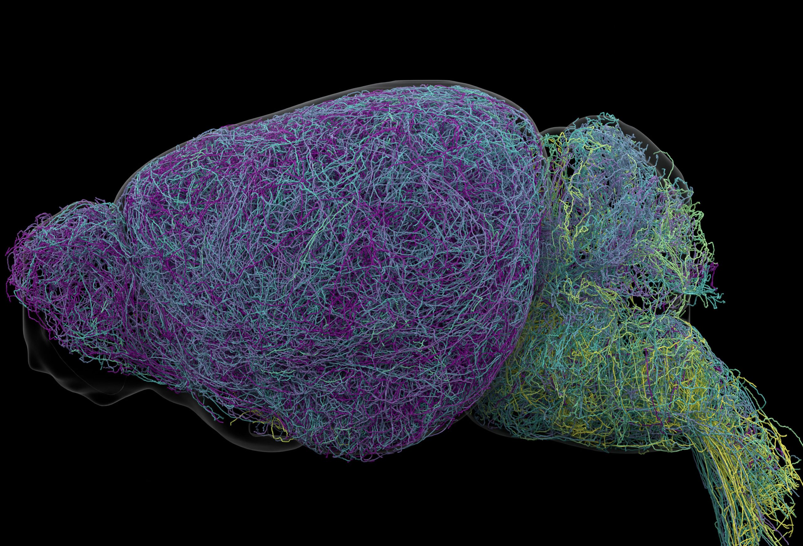
Scientists exploring the brain’s “blue place” discovered the longest neuron ever measured in a mouse and uncovered new clues about how the brain learns.
When mice in a decision-making experiment switched their choices after a negative outcome, neurons near the top of a tiny brainstem region became active. Just before the mice ignored cues that might lead to a reward, neurons near the bottom fired more rapidly.
These neighboring groups belong to the locus coeruleus (LC), Latin for “blue place.” This small cluster accounts for a tiny fraction of the brain’s cells but releases norepinephrine (NE), a chemical messenger that helps regulate attention and learning, activates the body’s response to stress or excitement, and affects heart rate.
Scientists long viewed the LC as a single loudspeaker broadcasting a uniform signal throughout the brain. In a new study published in Nature, Allen Institute researchers combined brain imaging, neural recordings, genetic tools, and behavioral experiments in mice to examine how the region’s connections and activity relate to what an animal experiences.
“The findings suggest the brain’s norepinephrine system is far more like a targeted postal network than a foghorn,” said Karel Svoboda, director of Neural Dynamics at the Allen Institute and study co-author.
Separate routes for learning and engagement
Tracing those connections revealed distinct destinations. Neurons in the upper, or dorsal, LC connected mainly to the cerebral cortex and forebrain. Those in the lower, or ventral, LC connected to the brainstem and spinal cord. These routes determine where the neurons send the signals that prompt norepinephrine release.
The work included whole-brain imaging of almost 35,000 neurons, genetic profiling of nearly 400,000 cells to examine gene activity, and complete reconstructions of some of the brain’s most complex cells. The researchers could therefore compare the neurons’ physical connections with their genetic characteristics and activity during behavior.
“What emerged was a clear map: neurons in the dorsal LC that send signals upward to the cortex are involved in learning, whereas neurons in the ventral region projecting downward to the brainstem and spinal cord govern whether animals engage with their environment at all,” said Svoboda. “We also showed that these anatomical differences are mirrored by distinct gene expression patterns.”
A 27.7-inch axon skips brain regions
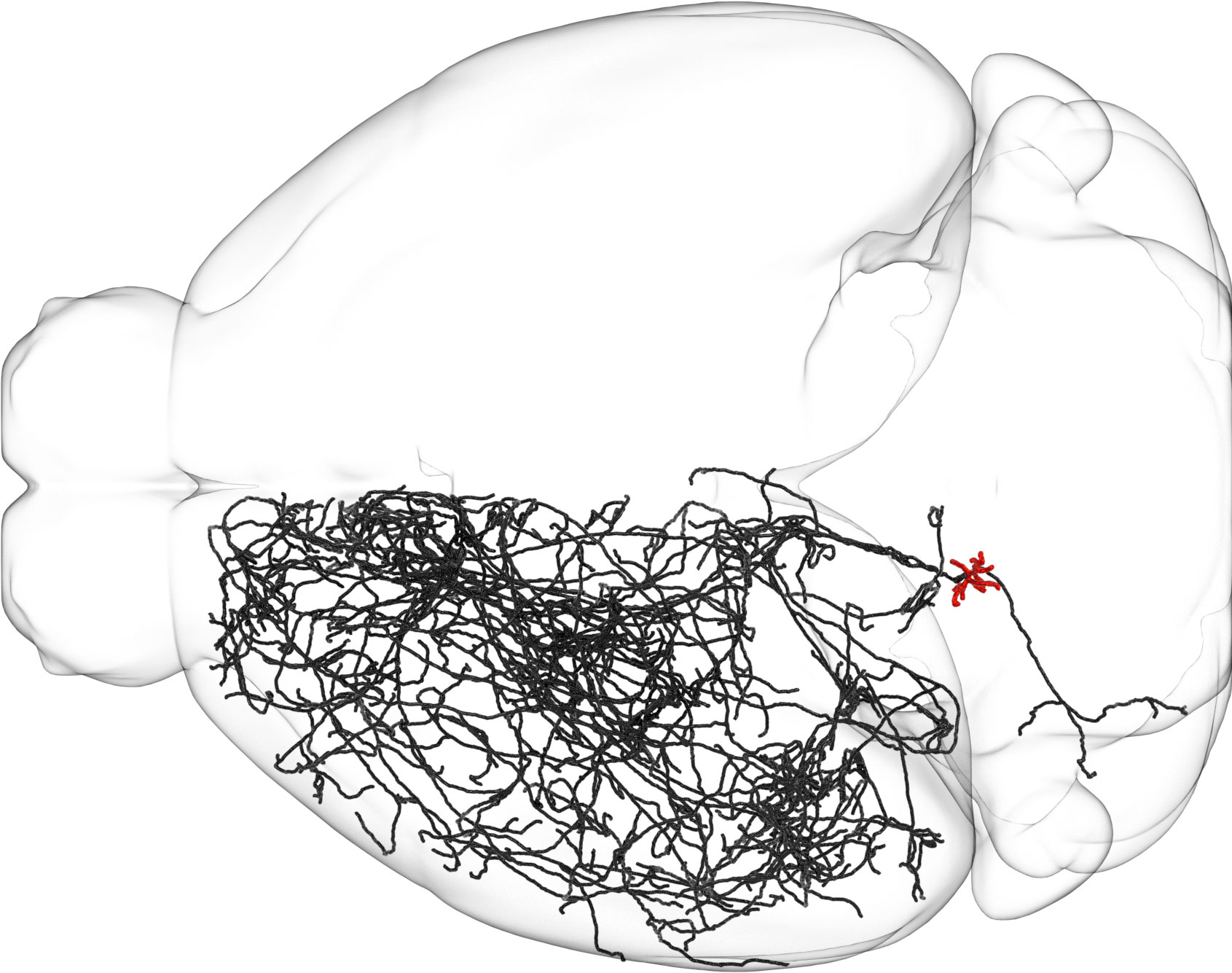
Reconstructing individual LC neurons also revealed how extensive their connections could be. Their axons, the long projections that carry signals to other cells, averaged about 35 centimeters, or 13.8 inches, in length. One measured 70.32 centimeters, about 27.7 inches, making it the longest single neuron ever measured in a mouse.
“This neuron, like many others that we studied, supplies NE to a very large volume of the cerebral cortex. For the brain, this is highly unusual. Most neurons are more specific in their targets,” said Jeremiah Cohen, a scientist at the Allen Institute and study co-author. “But this neuron doesn’t release NE everywhere. It ignores the cerebellum, brainstem, and spinal cord.”
The learning-related activity also led the researchers to draw parallels with dopamine, another major chemical messenger that carries signals to the basal ganglia, a brain region involved in habit formation. They propose that the norepinephrine and dopamine systems work together as a learning platform, allowing the brain to learn multiple complex and abstract concepts simultaneously.
Norepinephrine drugs could target specific circuits
Norepinephrine’s roles in mood and attention already make it a target of common medications for depression, anxiety, and ADHD. LC neurons are also among the first to degenerate in Alzheimer’s disease.
The organization uncovered in mice could eventually help researchers develop treatments that target particular parts of the system, reducing the possibility of helping some circuits while negatively affecting others.
Reference: “Topographic structure and function of locus coeruleus noradrenaline neurons” by Zhixiao Su, Polina Kosillo, Kanghoon Jung, Shuonan Chen, Mathew T. Summers, Alex Piet, Han Hou, Kenta M. Hagihara, Drew Friedmann, Olivia Ho-Shing, Matthew I. Becker, Thomas Chartrand, Xinxin Yin, Peter Grotz, Ella Hilton-VanOsdall, Margaret Lee, Rajvi Javeri, Samantha L. Tuggle, Naveen Ouellette, Holly Myers, Judith Baka, Camilo Laiton, Kaelin Wulf, John Rohde, Alessio P. Buccino, Cameron Arshadi, Di Wang, Sharmishtaa Seshamani, Sonya Vasquez, Carolyn M. Eng, Douglas R. Ollerenshaw, Nick Dee, Tamara Casper, Windy Ho, Matthew Jungert, Atlas Jordan, Elliot Phillips, Anish Bhaswanth Chakka, Kamiliam Nasirova, Krista Blake, Audrey McCutcheon, Megan Koch, Maria Camila Vergara, Kimberly A. Smith, Tim Jarsky, Nicholas Lusk, Mara C. P. Rue, Xiaoyin Chen, Joshua H. Siegle, Adam K. Glaser, Brian R. Lee, Karel Svoboda, Yoh Isogai, Jayaram V. Chandrashekar and Jeremiah Y. Cohen, 16 September 2026, Nature.
DOI: 10.1038/s41586-026-11026-0
Research reported in this publication was supported by the National Institutes of Health BRAIN Initiative under award numbers R01MH134833, R01NS104834 and RF1NS131984. This work was supported by the Allen Institute.
Never miss a breakthrough: Join the SciTechDaily newsletter.
Follow us on Google and Google News.