Scientists have captured an alarming new look at how Parkinson’s disease may begin. A toxic protein seems to drill tiny, flickering holes in brain cells, slowly wearing them down instead of destroying them outright.
Using a powerful imaging method, researchers watched this process unfold in real time, opening a fresh window into the hidden damage behind Parkinson’s symptoms and pointing to new ways to catch the disease earlier.
Parkinson’s Mystery Deepens
A newly identified toxic protein may hold the secret to how Parkinson’s disease develops. Scientists at Aarhus University report that this protein creates shifting pores in the membranes of brain cells, and with a new technique, they were able to watch these molecular attacks unfold in real time.
The first signs of Parkinson’s are usually easy to miss. A faint hand tremor. A little stiffness. Yet as time passes, brain cells begin to die and symptoms grow worse. For decades, the reason behind this decline has remained unclear, but researchers believe they are closer to finding an answer.
Protein Gone Rogue
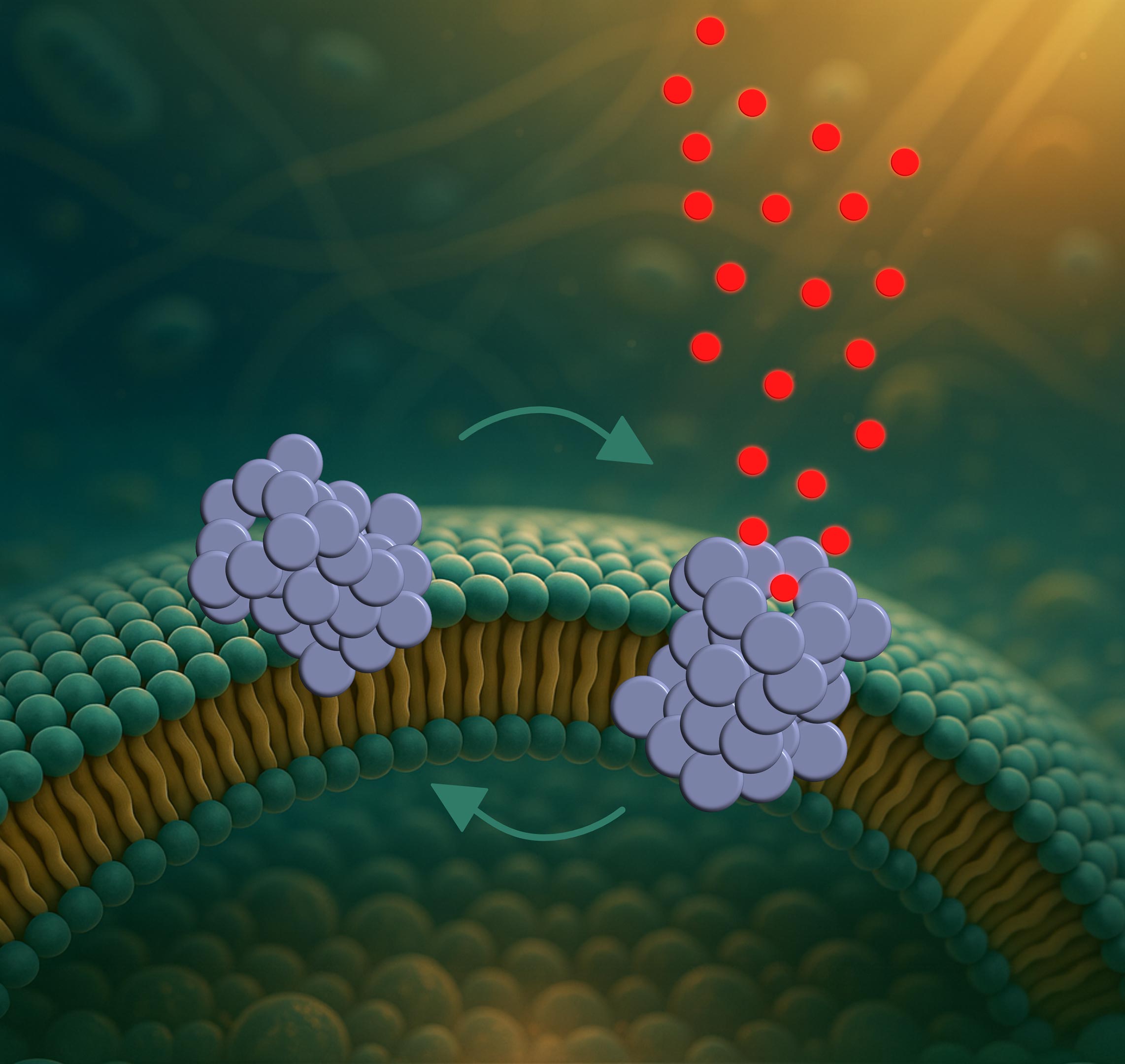
The focus is on a protein called α-synuclein. In a healthy brain, it helps nerve cells communicate with one another. In Parkinson’s disease, however, the protein starts to act differently, clumping together into harmful formations.
Most past studies have concentrated on large clusters of the protein, called fibrils, which can be seen in brain tissue from people with Parkinson’s. The new research, though, highlights much smaller and more dangerous shapes: α-synuclein oligomers. These tiny structures appear to puncture nerve cell membranes with microscopic holes.
The findings were published in ACS Nano, a journal of the American Chemical Society.
The video shows three artificial cell membranes being attacked by oligomers. The oligomers themselves are not visible, but their effect is. The membranes are colored blue and filled with small red fluorescent dyes. When the red colour disappears, it indicates that fluorescent dye has leaked out – evidence that the oligomers have created pores. The video is recorded at a resolution limit of 250 nanometer. Since the vesicles are 50–150 nanometer in diameter, the footage appears pixelated. Credit: Bo Volf Brøchner, AU
Tiny Revolving Doors in the Cells
“We are the first to directly observe how these oligomers form pores – and how the pores behave,” says Mette Galsgaard Malle, postdoctoral researcher at both Aarhus University and Harvard University.
The process unfolds in three steps. First, the oligomers attach to the membrane, especially at curved regions. Then they partially insert themselves into the membrane. Finally, they form a pore that allows molecules to pass through and potentially disrupt the cell’s internal balance.
But these are not static holes. The pores constantly open and close like tiny revolving doors.
“This dynamic behavior may help explain why the cells don’t die immediately,” says Bo Volf Brøchner, PhD student and first author of the study. “If the pores remained open, the cells would likely collapse very quickly. But because they open and close, the cell’s own pumps might be able to temporarily compensate.”

Molecular Movie in Slow Motion
This is the first time such pore dynamics have been observed in real time. It was made possible by a newly developed single-vesicle analysis platform that allows researchers to follow interactions between individual proteins and individual vesicles.
Vesicles are small artificial bubbles that mimic cell membranes and serve as simplified models of real cells.
“It’s like watching a molecular movie in slow motion,” explains Mette Galsgaard Malle. “Not only can we see what happens – we can also test how different molecules affect the process. That makes the platform a valuable tool for drug screening.”
Long Road to Treatment
In fact, the team has already tested nanobodies – small antibody fragments – developed to specifically bind these oligomers. They show promise as highly selective diagnostic tools. However, as a treatment, there is still some way to go.
“The nanobodies did not block the pore formation,” says Bo Volf Brøchner. “But they may still help detect oligomers at very early stages of the disease. That’s crucial, since Parkinson’s is typically diagnosed only after significant neuronal damage has occurred.”
The study also shows that the pores are not formed randomly. They tend to emerge in specific membrane types – especially those resembling the membranes of mitochondria, the cell’s energy factories. This could indicate that the damage begins there.
One Step at a Time
However, the researchers emphasize that the study was conducted in model systems – not in living cells. The next step will be to replicate the findings in biological tissue, where more complex factors come into play.
“We created a clean experimental setup where we can measure one thing at a time. That’s the strength of this platform,” says Mette Galsgaard Malle. “But now we need to take the next step and investigate what happens in more complex biological systems.”
Reference: “Single-vesicle Tracking of α-Synuclein Oligomers Reveals Pore Formation by a Three-Stage Model” by Bo Volf Bro̷chner, Xialin Zhang, Janni Nielsen, Jo̷rgen Kjems, Daniel E. Otzen and Mette Galsgaard Malle, 12 August 2025, ACS Nano.
DOI: 10.1021/acsnano.5c04005
The work was initiated and supervised by Professor Daniel E. Otzen (iNANO/MBG) together with Professor Jørgen Kjems (iNANO/MBG).
Never miss a breakthrough: Join the SciTechDaily newsletter.
Follow us on Google and Google News.
1 Comment
replenishing the blood cells in the brain is linear with brain function so a medicine for this would have to be a sleep medicine because the brain works in a pattern while in deep sleep. citric acid and fumes from chloride with a blood cell booster