
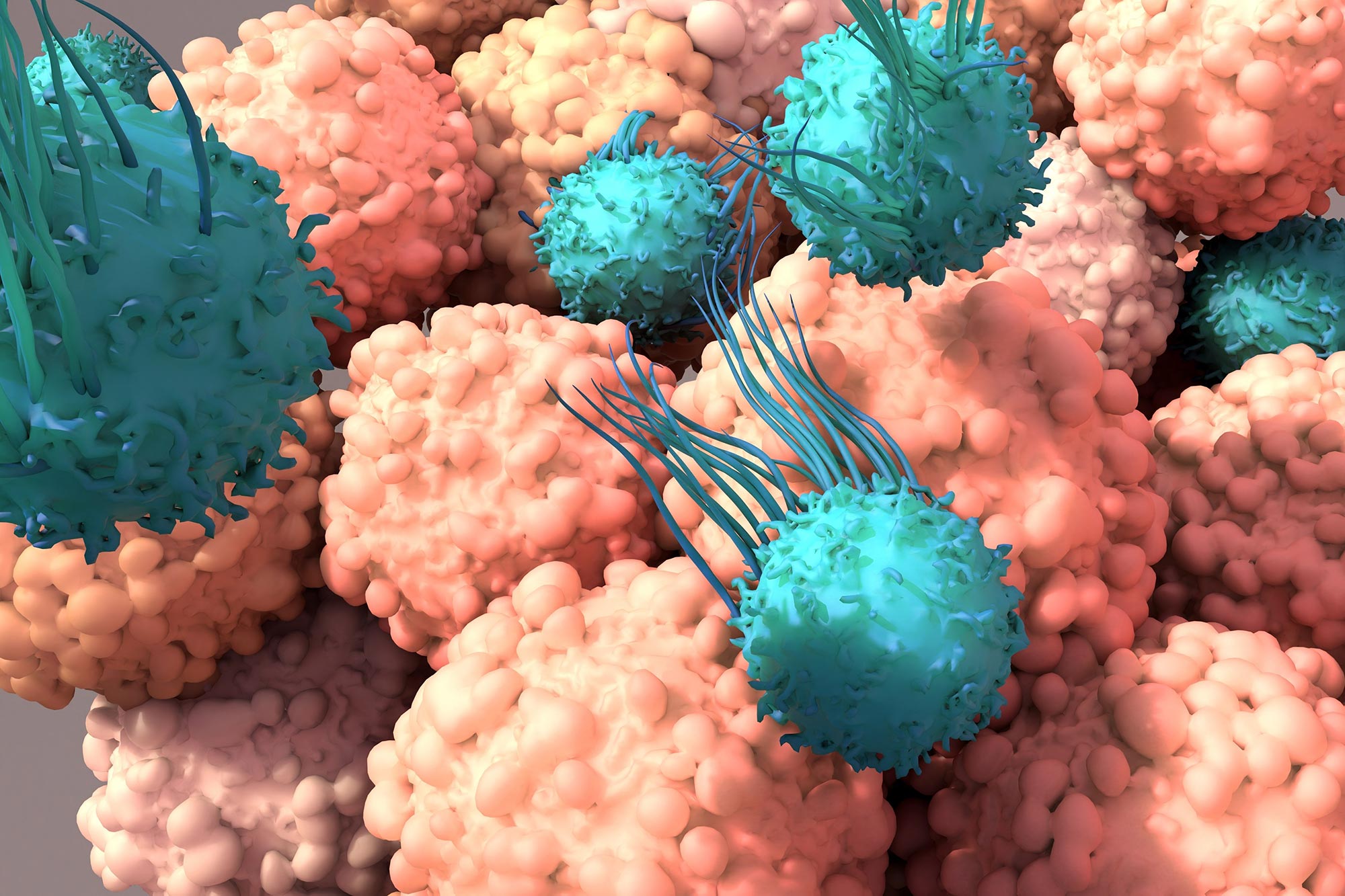
A study suggests that exhausted T cells may be burning energy too aggressively rather than simply running out of it.
Cancer immunotherapy can activate T cells, the immune system’s specialized killers, and direct them against tumors. Yet these cells often lose momentum before the cancer is controlled. Once they enter a state known as T cell exhaustion, they can no longer sustain a strong attack.
This loss of function has been a major limitation of checkpoint inhibitors, immunotherapy drugs that remove molecular brakes from T cells so they can fight cancer more aggressively.
“A tragic part of T cell exhaustion is that the immunotherapy seems to be working for patients, and then it fades,” says Santosha Vardhana, MD, PhD, a physician-scientist at Memorial Sloan Kettering Cancer Center (MSK) who treats people with lymphoma. “Many of them experience a brief wisp of promise only to have it taken away.”
Dr. Vardhana’s laboratory has now identified MEK, a signaling molecule, as an important driver of T cell exhaustion. The animal study, published in Immunity, indicates that inhibiting MEK could help prevent some T cells from becoming fully exhausted and potentially strengthen immunotherapy.
“We’re excited about applying this finding to enhance multiple forms of immunotherapy,” Dr. Vardhana says. “FDA-approved MEK inhibitors are already available, so this approach could be tested in humans without much delay.”
Exhaustion begins with an energy imbalance
Researchers only recently began to understand what pushes T cells into exhaustion. In 2020, Dr. Vardhana’s laboratory identified metabolism, the chemical processes cells use to obtain energy from nutrients, as an important starting point.
Repeated exposure to tumor antigens (the cancer proteins the immune system sees as foreign) places heavy demands on mitochondria, the structures inside T cells that convert nutrients into usable energy.
“There is a large metabolic demand being imposed as T cells encounter cancer cells and try to produce cancer-killing, or cytotoxic, proteins,” Dr. Vardhana says. “It turns out that the decision to make high levels of these proteins is regulated by MEK.”
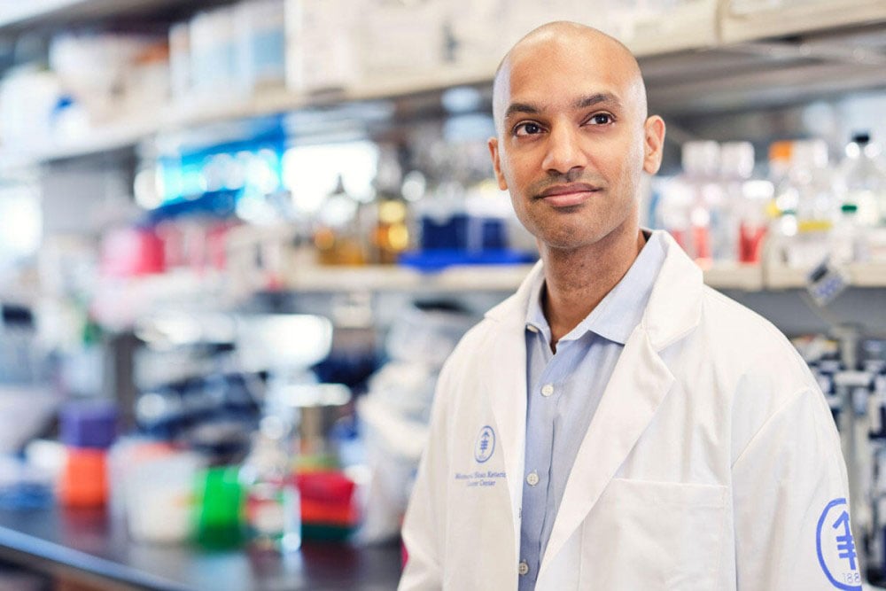
Excessive MEK activity eventually pushes T cells into terminal exhaustion, leaving them so depleted that immunotherapy can no longer reactivate them.
“We realized T cell exhaustion isn’t simply a loss of function — it reflects an imbalance between what these cells are being asked to do and the energy they have available,” says Tanmana Mitra, PhD, a student in the Vardhana lab and the study’s first author.
Exhausted cells spend energy on proteins
Despite appearing depleted, exhausted T cells remained highly active metabolically. When the researchers exposed them to MEK inhibitors, the cells multiplied more while consuming less energy.
“That paradox made us ask where all that energy was going, and we discovered that these cells were investing enormous resources into making proteins,” Dr. Mitra explains. “It changed how we think about T cell exhaustion — from a problem of too little energy to one of excessive energy demand.”

Reducing MEK signaling slowed the production of cytotoxic proteins, allowing some T cells to remain active and capable of renewing themselves for longer. This prolonged persistence could potentially improve the durability of immunotherapy.
The strategy resembles pacing a long drive instead of traveling at maximum speed from the beginning. Reducing the immediate intensity preserves enough capacity to continue much farther.
In laboratory models, suppressing MEK signaling helped T cells persist even within the difficult conditions surrounding a tumor.
Blocking MEK creates a tradeoff
MEK inhibition is unlikely to be equally useful for every cancer patient.
T cell exhaustion is more complicated than simply becoming weak. MSK immunologist Andrea Schietinger, PhD, found that exhaustion can protect T cells from overstimulation and death. By reducing their attack, the cells preserve themselves.
“As we’ve learned more about T cell exhaustion,” Dr. Vardhana says, “we’ve increasingly understood that it’s not the case that exhausted T cells are bad, so let’s try to reverse the process with a drug. Instead, exhaustion is more of an equilibrium state that lets the cells survive and keep going — almost like a ‘safe mode’ for T cells.”
When T cells attack and produce cytotoxic proteins, their mitochondria must convert nutrients into adenosine triphosphate (ATP). All living cells use ATP as their primary molecule for storing and transferring energy.
“Think of ATP as the currency in a fund that the cell spends down,” Dr. Vardhana says. “If you spend ATP on one thing, you don’t have enough to do something else. The exhaustion program is a sign that the cell’s bank account is getting close to zero. MEK tells exhausted cells whether to conserve fuel or go for broke. What we found is that inhibiting MEK makes the cells more conservative — helping them live longer while reducing the rate at which they produce the proteins that actually kill cancer cells.”
MEK therefore contributes to both the strength and the eventual failure of the immune response. It helps T cells attack at full power, but that intensity can lead to burnout. Inhibiting MEK preserves the cells while weakening their immediate cancer-killing activity.
Whether a brief, powerful attack or a slower, sustained response works better may depend on the characteristics of an individual patient’s cancer.
Patient needs may determine the strategy
Dr. Vardhana says MEK inhibitors should be used selectively. Two characteristics generally indicate that a patient is more likely to respond well to immunotherapy:
- The tumors are small.
- The patients have a high number of immune cells attacking the tumor — usually because the tumor has many mutations that make it recognizable.
“In these patients, conservation of T cells is not that important,” he says. “It’s like being in a car with 1/8 of a tank left, but you can see the finish line. In these patients, you would just let the car keep burning the gas — in other words, take the traditional immunotherapy approach. These are the patients in whom MEK inhibition is probably not needed.”
Patients with large tumors or relatively few immune cells may not produce a response powerful enough to eliminate the cancer quickly. In these cases, the slower response created by a MEK inhibitor may allow T cells to survive longer, which could be important when the tumor is large or the number of available T cells is limited.
Dr. Vardhana says carefully applied MEK inhibition could potentially strengthen several forms of immunotherapy:
- Checkpoint inhibitors: MEK inhibition has already been effective in treating melanoma when given along with checkpoint inhibitors and a targeted therapy called a BRAF inhibitor.
- Chimeric antigen receptor (CAR) T cell therapy: “We think this approach could dramatically boost T cell persistence, which has been a big problem with CAR T cell therapy,” Dr. Vardhana says.
- Tumor infiltrating lymphocyte (TIL) therapy: TIL therapy collects and expands immune cells that have already attacked the tumor. Using a MEK inhibitor before or after treatment may help the strongest tumor-fighting TILs survive longer.
- Bispecific antibodies: These laboratory-made proteins bind two targets at once. They can strongly activate T cells, but that increased activity may also drive exhaustion.
“This study shows the importance of understanding core principles of T cell biology — what sets the balance between conservation of energy and strong, cancer-fighting activity,” Dr. Vardhana says. “Once we know the answer to that, the therapeutic possibilities really start to fan out.”
Reference: “MEK-dependent bioenergetic demand drives terminal CD8+ T cell exhaustion” by Tanmana Mitra, Jahan Rahman, Madeline Hwee, Yan-Ting Chen, Ruben Jose Jesus Faustino Ramos, Hui Liu, Travis Hartman, Justin Cross, Miguel de Jesus, Morgan Huse, Valerie Longo, Pat Zanzonico and Santosha A. Vardhana, 13 July 2026, Immunity.
DOI: 10.1016/j.immuni.2026.06.012
The study was supported by an NCI K08 Career Development Award (NCI K08 CA237731), a Burroughs Wellcome Fund Career Award for Medical Scientists, a V Foundation Scholar Award, and the Josie Robertson Investigators program. This work was additionally supported by a Cancer Center Support Grant (P30 CA008748) and the Dorris J. Hutchison Pre-doctoral Fellowship by Memorial Sloan Kettering.
Never miss a breakthrough: Join the SciTechDaily newsletter.
Follow us on Google and Google News.