
A massive, multi-year project led by over 150 scientists has produced the most detailed map yet of how visual information travels through the brain – revealing more than 500 million connections in a speck of mouse brain tissue.
Using glowing neurons, high-powered electron microscopes, and deep learning, researchers captured both the physical wiring and real-time electrical activity of over 200,000 brain cells. The resulting 1.6-petabyte dataset is not just a technological marvel – it brings us closer to answering age-old questions about how our brains turn light into vision and how brain disorders might arise when this system breaks.
Unraveling the Brain’s Visual Code
In a major research effort funded by the National Institutes of Health (NIH), scientists have mapped the connections between hundreds of thousands of neurons in the mouse brain and recorded how they respond to visual input. By combining the brain’s wiring with real-time activity, this work lays crucial groundwork for understanding how the brain processes visual information to create the images we see.
The human brain processes information through the rapid electrical firing of about 86 billion neurons, each forming part of a vast network with trillions of connections. How we think, feel, and act is rooted in the structure of these connections and the electrical signals flowing through them in milliseconds. Although this new research focuses on just a tiny portion of the brain, it reveals how individual neurons are linked and how those links contribute to function. Insights like these could help explain how the brain works in health, and what goes wrong in conditions like injury or neurological disease.
Capturing the Brain in Action
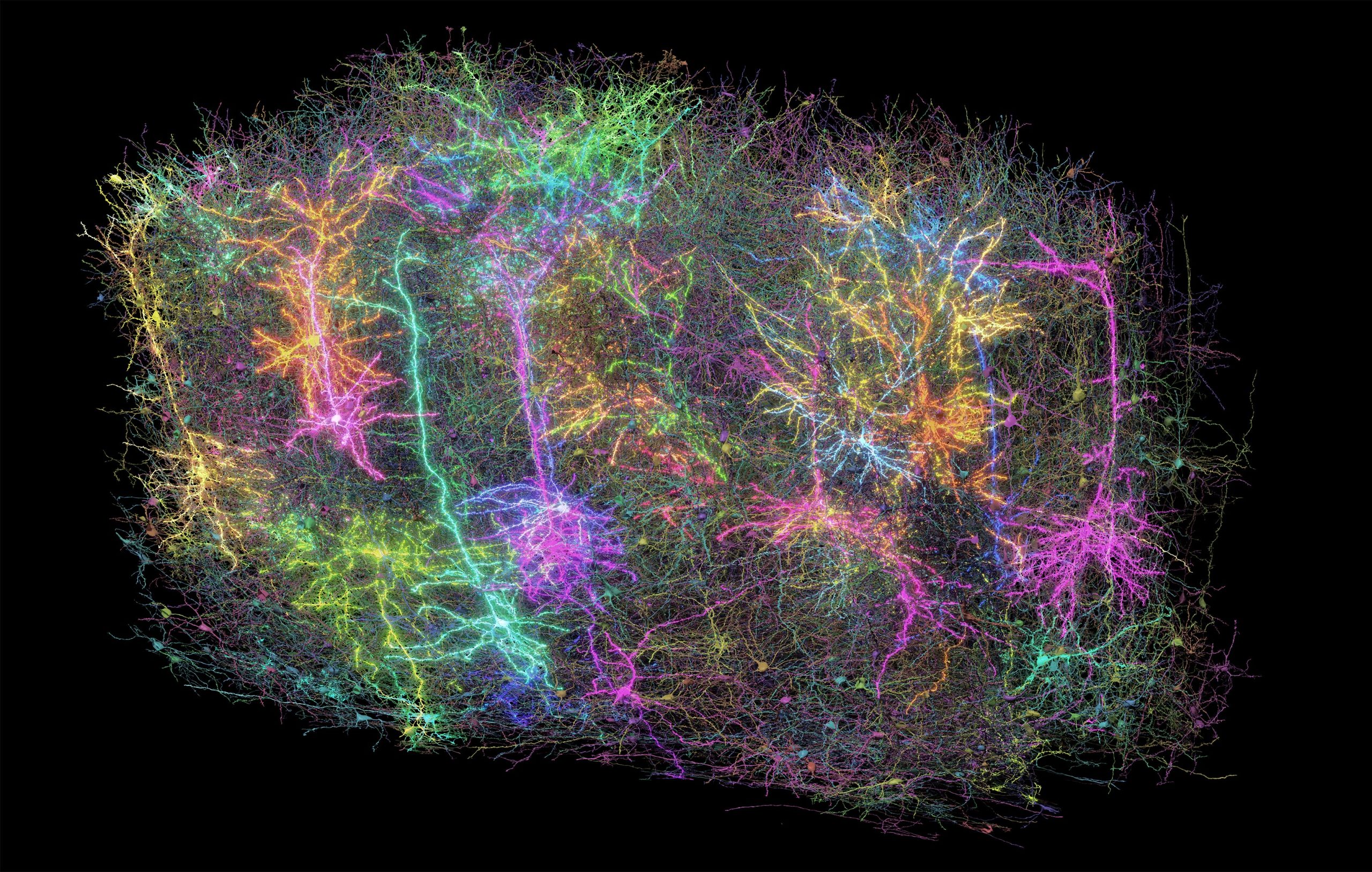
To conduct the study, researchers showed video clips to mice that had been genetically modified so that their neurons emit light when active. This allowed the team to record patterns of neuron activity in visual areas of the brain, covering a volume roughly the size of a grain of sand. Despite its small size, the tissue contained astonishing complexity: about four kilometers of axons – slender fibers that carry signals between neurons—interwoven to form over 524 million synapses among more than 200,000 cells.
Building the Brain’s 3D Circuit Map
To map these connections teams worked 12-hour shifts for 12 straight days to carefully cut and image ultra-thin slices of the brain tissue using electron microscopes (EM). Reconstruction was the most challenging next step, as it required accurately stitching together almost 28,000 EM images to align the connections that cross the volume of brain tissue.
This was followed by months of tracing the connections using deep learning algorithms followed by manual, and automated proofreading. Deep learning predictive models that explain visual information processing in the cortex were constructed and validated. In total, the sheer amount of data collected to create this tiny map comes out to 1.6 petabytes, roughly the equivalent of 22 years of continuous HD video.
A New Era of Neural Mapping
These results come at a time when maps of neurons and their connections are increasingly revealing the mysteries of the brain. In 2023, research funded by the National Institutes of Health Brain Research Through Advancing Innovative Neurotechnologies® Initiative, or The NIH BRAIN Initiative®, produced the first complete cell atlas of the mouse brain, including the types and locations surveyed from more than 32 million cells. Last year, the NIH BRAIN Initiative “Flywire” project led to the complete mapping of the common fruit fly brain, demonstrating the unique value of mapping the whole brain in its entirety.
The mouse connectome data detailed in this press release can be visualized online using the MICrONS Explorer resource.
Explore Further: A Grain of Brain, 523 Million Synapses, and the Most Complicated Neuroscience Experiment Ever Attempted
Reference: “Inhibitory specificity from a connectomic census of mouse visual cortex” by Casey M. Schneider-Mizell, Agnes L. Bodor, Derrick Brittain, JoAnn Buchanan, Daniel J. Bumbarger, Leila Elabbady, Clare Gamlin, Daniel Kapner, Sam Kinn, Gayathri Mahalingam, Sharmishtaa Seshamani, Shelby Suckow, Marc Takeno, Russel Torres, Wenjing Yin, Sven Dorkenwald, J. Alexander Bae, Manuel A. Castro, Akhilesh Halageri, Zhen Jia, Chris Jordan, Nico Kemnitz, Kisuk Lee, Kai Li, Ran Lu, Thomas Macrina, Eric Mitchell, Shanka Subhra Mondal, Shang Mu, Barak Nehoran, Sergiy Popovych, William Silversmith, Nicholas L. Turner, William Wong, Jingpeng Wu, Jacob Reimer, Andreas S. Tolias, H. Sebastian Seung, R. Clay Reid, Forrest Collman and Nuno Maçarico da Costa, 9 April 2025, Nature.
DOI: 10.1038/s41586-024-07780-8
Funding for this project was provided through the Machine Intelligence from Cortical Networks (MICrONS) Program of the Intelligence Advanced Research Projects Activity and the NIH BRAIN Initiative. The findings, published in a package of 10 papers published in the Nature family of journals, represent more than seven years of work performed by more than 150 scientists around the world.
Never miss a breakthrough: Join the SciTechDaily newsletter.
Follow us on Google and Google News.