Improved measurement of tissue stiffness could lead to better treatments for various conditions, including cancer and sports injuries.
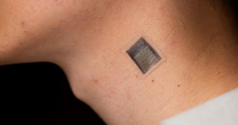
A group of engineers at the University of California San Diego has created a stretchable ultrasonic array that can perform non-invasive, serial 3D imaging of tissues as deep as 4 centimeters (1.6 inches) below the surface of the human skin. This innovative method boasts a spatial resolution of 0.5 millimeters and offers a more extended, non-invasive solution compared to current techniques, with enhanced penetration depth.
The study comes from the laboratory of Sheng Xu, a Professor of Nanoengineering at UC San Diego’s Jacobs School of Engineering and the lead author of the research. The findings were recently published in the journal Nature Biomedical Engineering.
“We invented a wearable device that can frequently evaluate the stiffness of human tissue,” said Hongjie Hu, a postdoctoral researcher in the Xu group and study coauthor. “In particular, we integrated an array of ultrasound elements into a soft elastomer matrix and used wavy serpentine stretchable electrodes to connect these elements, enabling the device to conform to human skin for serial assessment of tissue stiffness.”
Medical Applications of Continuous Elastography
The elastography monitoring system can provide serial, non-invasive, and three-dimensional mapping of mechanical properties for deep tissues. This has several key applications:
- In medical research, serial data on pathological tissues can provide crucial information on the progression of diseases such as cancer, which normally causes cells to stiffen.
- Monitoring muscles, tendons, and ligaments can help diagnose and treat sports injuries.
- Current treatments for liver and cardiovascular illnesses, along with some chemotherapy agents, may affect tissue stiffness. Continuous elastography could help assess the efficacy and delivery of these medications. This might aid in creating novel treatments.
In addition to monitoring cancerous tissues, this technology can also be applied in other scenarios:
- Monitoring of fibrosis and cirrhosis of the liver. By using this technology to evaluate the severity of liver fibrosis, medical professionals can accurately track the progression of the disease and determine the most appropriate course of treatment.
- Assessing musculoskeletal disorders such as tendonitis, tennis elbow, and carpal tunnel syndrome. By monitoring changes in tissue stiffness, this technology can provide valuable insight into the progression of these conditions, allowing doctors to develop individualized treatment plans for their patients.
- Diagnosis and monitoring for myocardial ischemia. By monitoring arterial wall elasticity, doctors can identify early signs of the condition and make timely interventions to prevent further damage.
Wearable Ultrasound
Wearable ultrasound patches accomplish the detection function of traditional ultrasound and also break through the limitations of traditional ultrasound technology, such as one-time testing, testing only within hospitals and the need for staff operation.
“This allows patients to continuously monitor their health status anytime, anywhere,” said Hu.
This could help reduce misdiagnoses and fatalities, as well as significantly cutting costs by providing a non-invasive and low-cost alternative to traditional diagnostic procedures.
“This new wave of wearable ultrasound technology is driving a transformation in the healthcare monitoring field, improving patient outcomes, reducing healthcare costs and promoting the widespread adoption of point-of-care diagnosis,” said Yuxiang Ma, a visiting student in the Xu group and study coauthor. “As this technology continues to develop, it is likely that we will see even more significant advances in the field of medical imaging and healthcare monitoring.”
The array conforms to human skin and acoustically couples with it, allowing for accurate elastographic imaging validated with magnetic resonance elastography.
In testing, the device was used to map three-dimensional distributions of the Young’s modulus of tissues ex vivo, to detect microstructural damage in the muscles of volunteers prior to the onset of soreness and monitor the dynamic recovery process of muscle injuries during physiotherapy.
The device consists of a 16 by 16 array. Each element is composed of a 1-3 composite element and a backing layer made from a silver-epoxy composite designed to absorb excessive vibration, broadening the bandwidth and improving axial resolution.
Reference: “Stretchable ultrasonic arrays for the three-dimensional mapping of the modulus of deep tissue” by Hongjie Hu, Yuxiang Ma, Xiaoxiang Gao, Dawei Song, Mohan Li, Hao Huang, Xuejun Qian, Ray Wu, Keren Shi, Hong Ding, Muyang Lin, Xiangjun Chen, Wenbo Zhao, Baiyan Qi, Sai Zhou, Ruimin Chen, Yue Gu, Yimu Chen, Yusheng Lei, Chonghe Wang, Chunfeng Wang, Yitian Tong, Haotian Cui, Abdulhameed Abdal, Yangzhi Zhu, Xinyu Tian, Zhaoxin Chen, Chengchangfeng Lu, Xinyi Yang, Jing Mu, Zhiyuan Lou, Mohammad Eghtedari, Qifa Zhou, Assad Oberai and Sheng Xu, 1 May 2023, Nature Biomedical Engineering.
DOI: 10.1038/s41551-023-01038-w
The study was funded by the Air Force Research Laboratory and the National Institutes of Health.
Professor Xu is now commercializing this technology via Softsonics LLC.
Never miss a breakthrough: Join the SciTechDaily newsletter.
Follow us on Google and Google News.