

The newly identified lymphatic network could fundamentally change how scientists understand and treat some of the leading causes of blindness.
Fluid, proteins, and inflammatory debris can accumulate behind the retina in several major causes of permanent blindness, including glaucoma and age-related macular degeneration. Yet scientists have long struggled to explain how the eye normally removes this material.
Researchers at the University of British Columbia and the University of Toronto have now traced what appears to be a previously unknown drainage route at the back of the eye. Called the posterior ocular lymphatic outflow, or POLO pathway, it carries fluid and waste out of the eye and into the body’s lymphatic system.
“The retina is one of the most metabolically active parts of the body, constantly generating byproducts that need to be cleared,” said Dr. Neeru Gupta, professor and head of UBC’s department of ophthalmology and visual sciences. “This discovery helps explain how the eye flushes this waste and promises to transform how we think about and treat a range of eye conditions.”
A cleanup route connects eye disease
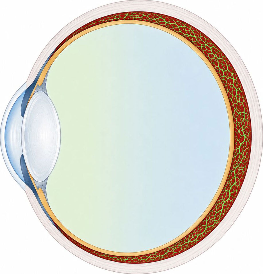
Glaucoma, macular degeneration and other retinal disorders are associated with excess fluid, metabolic waste, tissue stress and inflammation. Age-related macular degeneration alone affects about 2.5 million people in Canada.
The identification of the POLO pathway suggests that the eye has a natural mechanism for removing this material. Researchers say the route could eventually become a target for treatments designed to improve clearance.
“This gives us a whole new framework for understanding these diseases and a potential target for therapeutics,” said Dr. Gupta. “The question now is: How can we enhance or exploit this cleanup system to treat or even prevent disease.”
A century-old assumption begins to fall
For more than 100 years, the eye was generally believed to have no lymphatic system. In most other organs, lymphatic vessels help control fluid levels, remove waste and support immune activity.
Dr. Gupta and Dr. Yeni Yücel, professor and director of ophthalmic pathology at the University of Toronto, began questioning that view in 2009. They identified a lymphatic-related drainage route at the front of the eye known as the “uveolymphatic” pathway.
The back of the eye remained much less understood, even though it contains the retina and is where many severe forms of vision loss begin.
“The retina is responsible for vision, and it’s also where many of the most serious vision-loss diseases occur,” said Dr. Yücel. “Understanding how this part of the eye maintains a balanced environment and flow of materials is critical.”
Imaging traces waste beyond the eye
To search for the POLO pathway, the researchers studied mice using MRI, near-infrared fluorescence and microscopic analysis. They placed fluorescent tracer molecules into a narrow space behind the eye and followed their movement in real time.
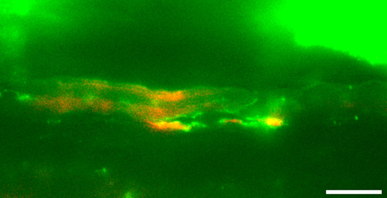
The tracers revealed small lymphatic vessels within the choroid, a thin layer of tissue located beneath the retina. Fluid moved from the back of the eye into nearby orbital tissue and reached surrounding lymph nodes within minutes, connecting the eye with the larger lymphatic network.
“Because lymphatic vessels in the choroid were thought not to exist, the team used multiple techniques to demonstrate both their presence and function,” added Dr. Yücel, a pathologist-scientist at St. Michael’s Hospital. “We were surprised to see such a direct route for fluid to leave the eye and connect with the lymphatic system. It suggests the back of the eye has an active clearance pathway, which could play an important role in removing fluid, proteins and inflammatory material that build up in disease.”
Human therapies remain a future goal
Researchers still need to determine whether the pathway functions the same way in people and whether it can be safely influenced with treatments.
In the future, the discovery could support more effective delivery of drugs to the back of the eye, therapies that improve fluid removal and a better understanding of how pressure, inflammation and fluid movement contribute to vision loss.
“This is a foundational discovery that shows the eye is not as closed a system as we previously thought,” said Dr. Gupta. “It gives us a new map, a new mechanism and a new set of questions to explore.”
Reference: “Identification and Mapping of a Posterior Ocular Lymphatic Outflow (POLO) Pathway Through Choroidal Lymphatics” by Babishaa Saunthararajan, Neeru Gupta, Xun Zhou, Ekim Gümeler, Greg Stanisz, Wilfred W. Lam, Margaret Koletar and Yeni H. Yücel, July 2026, Translational Vision Science & Technology.
DOI: 10.1167/tvst.15.7.23
The research was supported by the Canadian Institutes of Health Research, Glaucoma Research Society of Canada, Henry Farrugia Ophthalmology Research Fund, Canadian Space Agency, Dorothy Pitts Chair, Stephen M. Drance Chair, Thor and Nicky Eaton Research Fund and Canada Foundation for Innovation Leaders Opportunity Fund.
Never miss a breakthrough: Join the SciTechDaily newsletter.
Follow us on Google and Google News.
3 Comments
I wonder if “floaters” could somehow be channeled out of the vitreous.
Yes,by way of a vitrectomy of course, yet this new discovery may be a way of the eye doing that itself?
Get an AI working on this. Humans are too slow!