
The discovery could offer new clues about chronic inflammatory conditions.
Cells in tissues such as the skin, gut, and airways regularly die and need to be removed. Their neighbors face a difficult task: they must swallow the dying cell without leaving a gap in the protective layer around it.
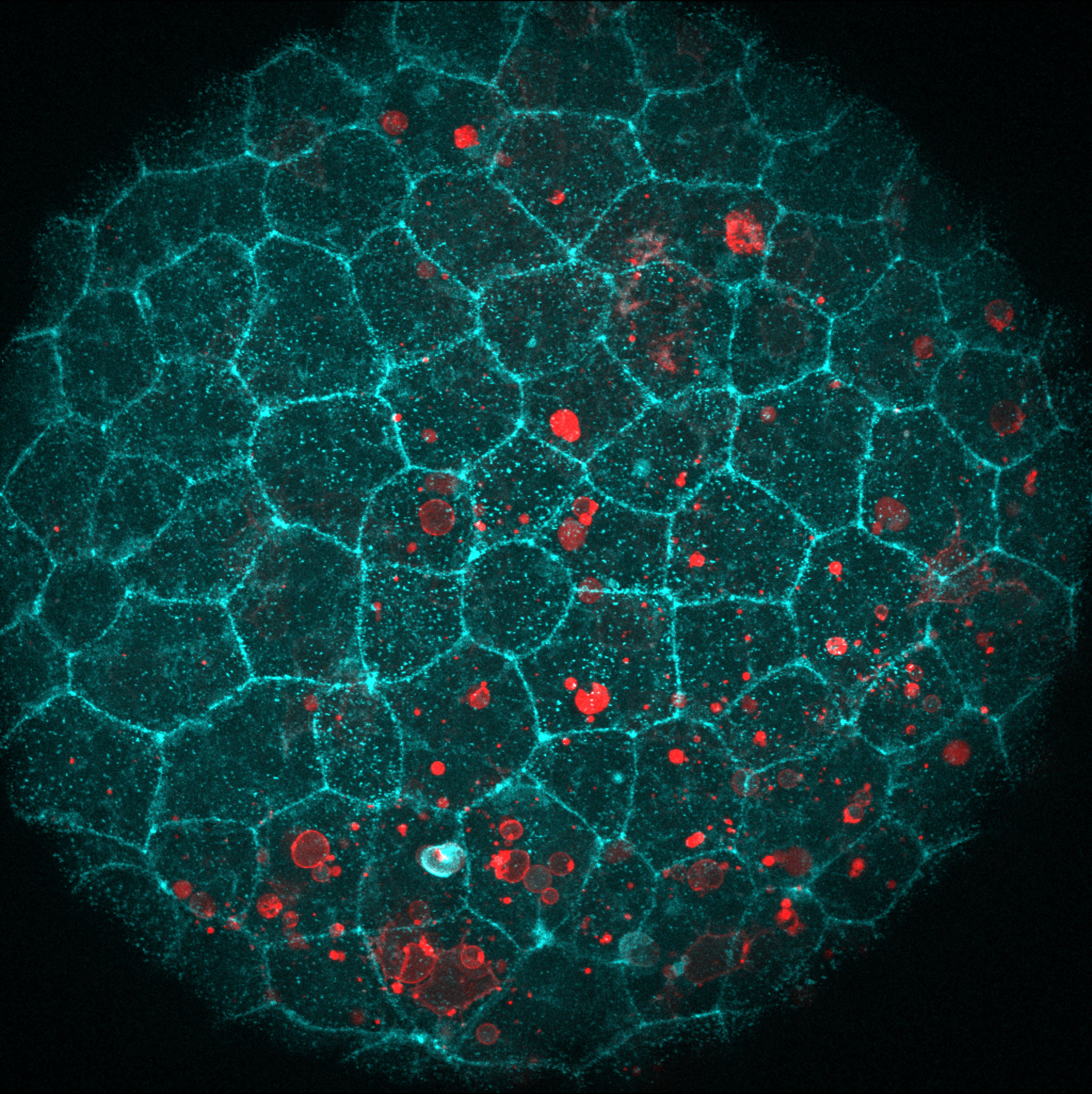
Researchers at the Center for Genomic Regulation in Barcelona have discovered how cells can accomplish both jobs at once. In zebrafish and mouse embryos, cells reused part of the molecular machinery that normally holds neighboring cells together to help remove dying cells while keeping the surrounding tissue sealed.
“We were intrigued to find out that epithelial cells repurpose their molecular adhesion machinery – the ‘glue’ that normally holds them together – to engulf dying cells,” says ICREA Research Professor Verena Ruprecht, senior author of the study.
Cellular glue doubles as cleanup machinery
The cells involved are called epithelial cells. They form tightly connected layers that cover and line surfaces throughout the body, including the skin, gut, and airways. One of the proteins helping these cells stick together is E-cadherin, which works with several other proteins as part of a larger molecular complex.
Live imaging showed that this machinery gathered precisely where an epithelial cell came into contact with a dying neighbor. The researchers then asked whether E-cadherin was simply attaching the two cells in the same way it normally connects healthy neighboring cells.
To find out, they first presented the tissue with dying cells that had been stripped of E-cadherin. The epithelial cells still removed them efficiently. The team then used droplets of fat that contained no proteins but carried a surface signal normally displayed by dying cells. The tissue swallowed those droplets as well.
Together, the experiments showed that E-cadherin on the dying cell was not necessary for removal. Instead, the E-cadherin complex inside the surrounding tissue appears to take on a different role, helping epithelial cells physically engulf the material they need to clear.
Cells swallow neighbors without breaking the barrier
Doing that without weakening the tissue creates another challenge. Epithelial cells are packed closely together to form a continuous barrier, but swallowing something about the size of another cell requires a major change in shape.
The researchers found that an epithelial cell solves this problem by allowing its two surfaces to behave differently. The lower surface stretches around the dying cell and helps pull it inside, while the upper surface remains relatively stable and keeps the tissue sealed.
Measurements from the live imaging supported this division of labor. The upper surface changed very little before, during, and after engulfment, while the lower surface underwent much larger changes in shape.
Ruprecht compares the behavior to dancers standing in a line with their arms linked. Their upper bodies remain steady while their feet perform the more complicated movement needed when a dying cell appears.
“It’s the same dancer with a different choreography,” she says.
Cell cleanup requires balanced force
The team also investigated how individual proteins in the E-cadherin complex contribute to the process. One component connects the complex to the cell’s internal skeleton, allowing the cell to transmit force as it wraps around and engulfs a dying neighbor.
When the researchers removed that protein, or the specific part needed to connect with the internal skeleton, the cells could no longer engulf dying cells.
Another protein helped restrain the cell’s contractile machinery. Removing that restraint did not make the cells better at engulfing their targets. Instead, the cells became too stiff and failed to clear the dying cells.
The researchers then tested whether the same mechanism operates beyond zebrafish. In early mouse embryos, blocking E-cadherin also left dying cells uncleared, providing evidence that the process is shared across vertebrates.
Embryos are especially useful for studying this kind of behavior because they are transparent, allowing researchers to watch living cells and tissues in detail as the cleanup occurs.
The study builds on Ruprecht’s earlier work showing that epithelial cells in embryos can cooperate to remove dying cells as a form of early innate immune defense. The new findings, published on August 27, 2026 in Nature Communications, help explain how those cells perform the physical cleanup without breaking the barrier they form.
Failed cleanup could fuel inflammation
Whether the same E-cadherin mechanism operates in adult zebrafish, adult mice, or human tissues remains unknown.
There are reasons to investigate further. Adult epithelial tissues in the retina, colon, airways, and mammary gland are already known to remove dying cells, and E-cadherin is found in epithelial tissues throughout the body. Its structure has also changed remarkably little across species.
If the mechanism proves to operate more broadly, it could help researchers better understand how tissues safely dispose of dying cells. Cells that are not removed can eventually break apart and release their contents, which can contribute to chronic inflammation.
The findings suggest that successful cleanup depends on more than recognizing the chemical signals given off by a dying cell. The surrounding tissue must also be able to reshape itself enough to remove that cell without compromising the protective barrier.
“Studying the mechanisms of how dying cells can be removed efficiently from tissues is of very high relevance to human health,” concludes Ruprecht.
Reference: “De novo E-cadherin/catenin complex formation controls basal epithelial mechanics and force transmission for apoptotic cell clearance” by Hanna-Maria Häkkinen, Marta Batet, Laura F. Bianchi, Senda Jiménez-Delgado, Fabio Pezzano, Stefan Wieser, Luca Ciampa, Esteban Hoijman, Carina Vibe, Sofie Wijma and Verena Ruprecht, 27 August 2026, Nature Communications.
DOI: 10.1038/s41467-026-76710-1
Funded by the Spanish Ministry of Science and Innovation, the Human Frontier Science Program, the European Union’s Horizon Europe program, and the “la Caixa” Foundation, with additional support from the European Social Fund. It made use of the CRG Core Facilities for Advanced Light Microscopy, Tissue Engineering, and Protein Technologies.
Never miss a breakthrough: Join the SciTechDaily newsletter.
Follow us on Google and Google News.