
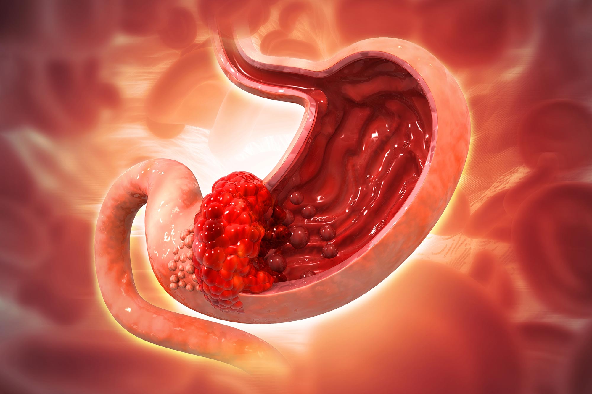
A common stomach bacterium may trigger a developmental repair program that pushes gastric tissue toward a precancerous state.
Stomach cancer often begins years before a tumor can be detected. During that long interval, the stomach lining can undergo a series of molecular and cellular changes that gradually push healthy tissue toward a precancerous state.
Scientists at Charité – Universitätsmedizin Berlin have now mapped a key part of that process in unprecedented detail. Their study shows how chronic infection with Helicobacter pylori, a major cause of stomach cancer, alters communication among immune cells, connective tissue, and the cells lining the stomach. The findings, published in Nature Communications, identify a chain of signals that may eventually provide new ways to interrupt cancer development before a tumor forms.
A Common Infection With Long Term Consequences
H. pylori is a widespread bacterium that can colonize the stomach for decades. It is often acquired within families and can cause chronic inflammation, ulcers, and, in some people, changes that increase the risk of stomach cancer.
That risk is particularly relevant for people with a family history of gastric cancer linked to H. pylori. Researchers led by Prof. Michael Sigal and Dr. Manqiang Lin at Charité are studying what happens in the stomach tissue of such patients long before cancer becomes detectable.
“We encounter precisely these kinds of people at the clinic, and they are often very anxious. If we understand what happens in the stomach tissue even before cancer develops, we might be able to intervene,” says Michael Sigal, professor of translational gastrointestinal oncology.
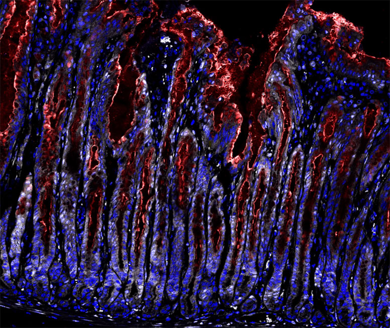
The Missing Link Between Inflammation and Tissue Remodeling
Scientists have known for decades that chronic H. pylori infection raises the risk of stomach cancer. What remained less clear was how infection-driven inflammation causes the stomach lining to change its identity and growth behavior.
“We knew from previous studies that the bacterium disturbs the balance of growth signals in the stomach lining.” But that did not explain an unusual observation. The infected mucosa begins growing rapidly even though the stomach’s classic stem cells are not expanding in the way researchers would normally expect.
“So, there must be another program at work that has been overlooked until now,” Sigal says.
To identify that program, the researchers analyzed tens of thousands of individual cells from diseased stomach tissue using single-cell sequencing. The technique allowed them to determine which genes were active in different cell types during infection.
They then used animal models in which specific genes could be selectively switched off in particular cells, allowing the team to test which signals were necessary for the tissue changes they observed.
Reconstructing the Cellular Conversation
The researchers also grew miniature versions of stomach tissue known as organoids. They combined these with connective tissue cells to create assembloids, experimental systems that allowed them to study how different cell populations communicate.
By selectively interrupting specific signaling pathways, the team could determine which cells were sending particular molecular messages and which cells were responding.
The researchers compared those results with publicly available human datasets to examine whether the same cellular patterns were also present in human stomach tissue.
Their experiments revealed a multistep process in which one cellular change prepares the way for the next. Over time, the sequence can contribute to precancerous lesions and, eventually, cancer.
“The infected stomach lining doesn’t just grow faster, but actually transitions into a fundamentally different state. A process is gradually activated that normally occurs only during embryonic development and wound healing,” explains first author Giulia Beccaceci, an early career researcher in Sigal’s group.
An Injury Repair Program Goes Off Course
The process begins when H. pylori disrupts the stomach lining’s normal defenses. A signal involved in regulating the renewal of surface cells and their tolerance of bacteria is lost.
The immune system then responds to the infection. Immune cells enter the affected tissue and release inflammatory molecules, particularly interleukin-1β.
Rather than acting primarily on the stomach lining itself, interleukin-1β affects the connective tissue beneath it. Those connective tissue cells then produce another signal that changes the behavior of the overlying mucosa.
A tissue hormone normally involved in wound healing switches the stomach lining into a repair program. Under persistent inflammatory conditions, however, that program does not simply restore damaged tissue. Cells begin dividing more frequently and the mucosa shifts into a different biological state.
“So connective tissue isn’t just a bystander, but the actual switch,” Beccaceci says.
The researchers conclude that the connective tissue acts as a signaling intermediary between inflammation and abnormal growth in the stomach lining. That triangular communication helps explain how a chronic bacterial infection can produce lasting changes in tissue architecture.
Stopping the Process Before Cancer Forms
Removing H. pylori remains one of the clearest strategies for lowering stomach cancer risk.
“Persons with an increased risk—for example, due to a family history of stomach cancer, symptoms, or a known infection—should be tested for the stomach bacterium and, if the test is positive, treated with antibiotics,” Sigal says. “This is simple and has been proven to reduce the risk of stomach cancer. This is due to the fact that it removes the persistent irritation from the tissue that keeps the disease-causing chain of changes in motion.”
Eradicating the bacterium does not always return the stomach lining completely to its original state. In some patients, the tissue appears to remain reprogrammed even after the infection is gone, leaving a residual cancer risk.
That problem has led the Charité team to search for molecular markers that could identify people whose stomach tissue has already entered a precancerous trajectory.
“In order to reliably identify these individuals, we are currently developing markers that can detect, in mucosal tissue samples, whether the tissue is on its way to becoming precancerous,” Sigal says. “In addition, we now understand the individual steps in the chain and, as a result, the potential targets for drugs.”
A New Target in the Inflammatory Pathway
One possible intervention would be to block the response of connective tissue cells to interleukin-1β. If those cells no longer reacted to the inflammatory signal, the downstream changes in the stomach lining might be prevented.
Previous population studies offer indirect support for targeting inflammatory pathways. Regular use of some common anti-inflammatory drugs has been associated with a lower risk of gastrointestinal tumors. These drugs can inhibit COX-2, an enzyme that participates in the newly described signaling network.
Prevention Before a Tumor Exists
Most cancer prevention and screening programs focus on detecting disease at an early stage. Sigal’s group is investigating an even earlier window, when tissue has begun moving toward a precancerous state but no tumor has yet developed.
Charité is establishing a Cancer Prevention Clinic to study and monitor people with elevated stomach cancer risk. Researchers working with the ImmunoPreCept Cluster of Excellence plan to use tissue samples from these patients to better define the molecular stages that precede cancer.
“While we now know that cancer can be prevented if interventions are taken early enough, it remains to be seen, however, whether a tissue that has already been reprogrammed can return to its normal state,” Sigal says.
Reference: “Helicobacter pylori triggers gastric mucosal remodeling toward a fetal-like transcriptional program via stromal IL-1β signaling” by Giulia Beccaceci, Stefanie Müllerke, Hilmar Berger, Christian Täger, Ronja Möbius, Anne-Sophie Fischer, Kimberly Hartl, Jonas Wizenty, Hans-Joachim Mollenkopf, Michael Naumann, Manqiang Lin and Michael Sigal, 5 September 2026, Nature Communications.
DOI: 10.1038/s41467-026-77520-1
The research was funded by, among others, the European Research Council (ERC Starting Grant REVERT), the Deutsche Forschungsgemeinschaft, the Federal Ministry of Research, Technology, and Space, the Einstein Center 3R, and the ImmunoPreCept Excellence Cluster.
Never miss a breakthrough: Join the SciTechDaily newsletter.
Follow us on Google and Google News.