

Researchers tracking glioblastoma cells in living mouse brain tissue discovered how quickly they can change their paths, revealing a hidden challenge in stopping this aggressive brain cancer.
Glioblastoma is one of the most aggressive forms of brain cancer, largely because its cells can spread deep into surrounding healthy tissue. Even after surgeons remove the main tumor, scattered cancer cells may remain in the brain and eventually contribute to the disease returning.
Understanding how these cells spread has been difficult because researchers have largely relied on images that capture only individual moments. Scientists at Uppsala University have now developed a method that lets them watch thousands of glioblastoma cells move through living brain tissue, revealing how they change their behavior over time. The findings, published in Nature Communications, could help researchers better understand why this cancer is so difficult to contain.
“Until now, we have mostly seen snapshots of how glioblastoma spreads. This method combines microscopy with artificial intelligence and allows us to watch the process unfold in living brain tissue. We could also compare tumors from different patients and investigate how they respond to treatment,” says Hitesh Mangukiya, researcher at the Department of Immunology, Genetics and Pathology at Uppsala University and one of the study’s lead authors.
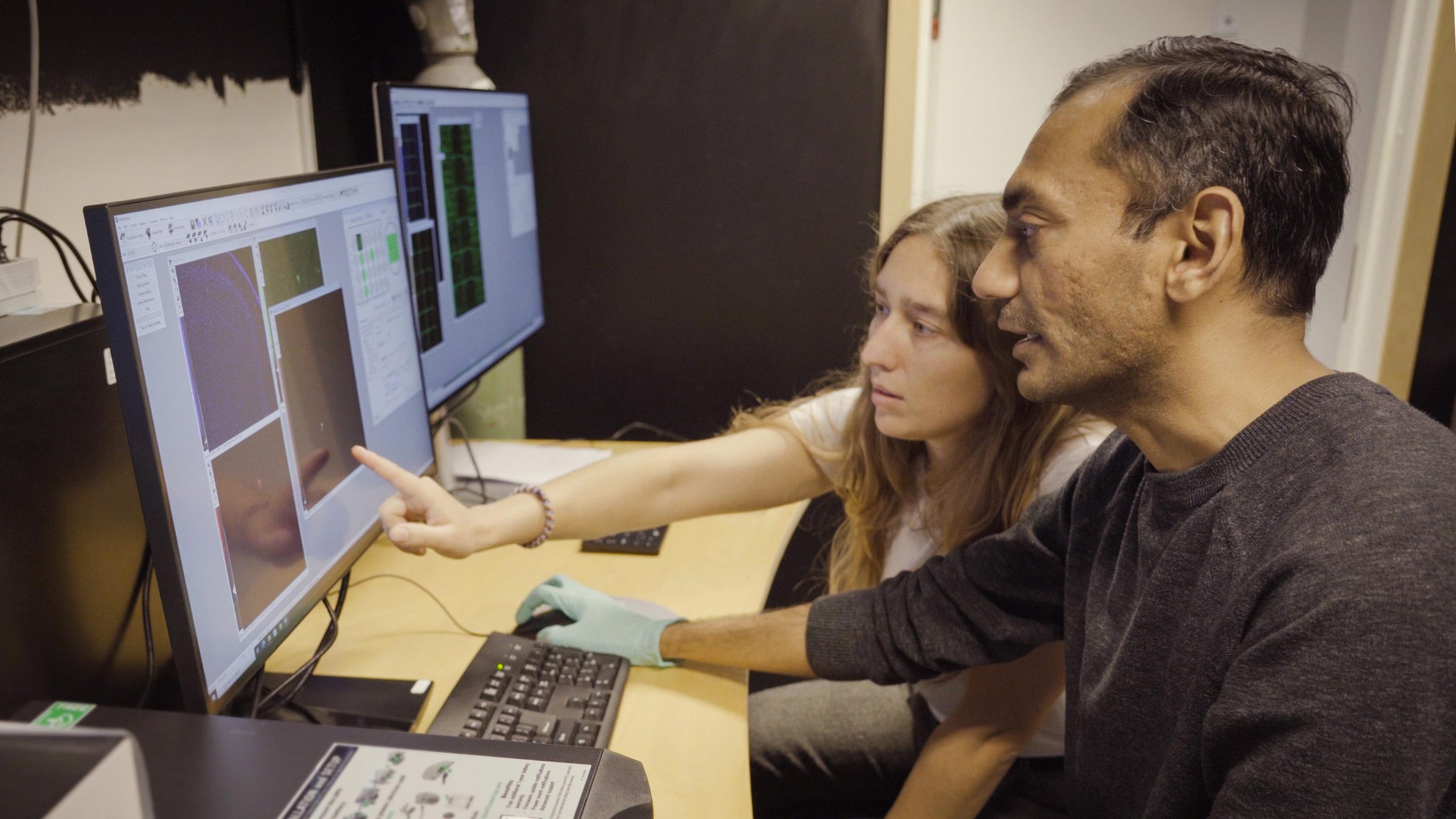
The new method, called GlioTrace, combines automated microscopy with machine learning to follow individual cancer cells as they travel through brain tissue. To test it, the researchers collected glioblastoma cells from six patients and grew them as tumors in mouse brains. They then cut the brains into thin slices, kept the tissue alive in the laboratory, and photographed it at intervals of no more than two hours for up to five days.
A computer system identified and tracked individual tumor cells across the images, while statistical models helped researchers calculate when cells switched between different states. Unlike techniques that examine only small regions of the brain, GlioTrace can monitor cells across entire brain slices using standard microscopes.
Cancer cells switch paths through the brain
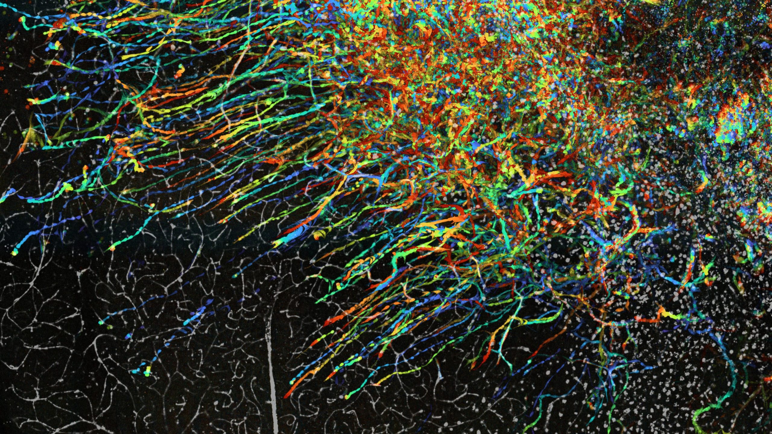
The observations revealed that glioblastoma cells do not all spread in the same way. Some tumors showed a preference for traveling along blood vessels, while others spread more diffusely through the surrounding brain tissue. These differences were apparent between tumors derived from different patients.
Individual cells were also remarkably adaptable. A cancer cell moving along a blood vessel could later leave that route and travel independently through the tissue. Some cells changed their shape and movement patterns over just a few days, suggesting that their behavior is far less fixed than individual images might imply.

“What surprised us most was how flexible the tumor cells are. The same cell can change how it moves over just a few days. To develop treatments that stop tumor cells from spreading, we need to understand this ability to switch between different behaviors,” says Madeleine Skeppås, PhD student at Uppsala University and co-lead author of the study.
Cancer treatments affect migrating cells differently
The team also used GlioTrace to examine how cancer treatments influence cell movement. Two drug substances reduced the movement and spread of tumor cells in the brain slices, although their effectiveness varied between patient-derived tumors. Even within the same research setting, cells with different movement patterns did not respond equally to the drugs.
Another experiment raised questions about what happens to invasive cells during treatment. In some tumor models, combining radiation with chemotherapy increased the proportion of cells with a particular shape. These cells were among the fastest moving and occupied a central position in the network of transitions between different cell states, indicating that they were closely connected to changes in cell behavior.
The findings suggest that certain highly invasive cells might persist during treatment. However, the researchers have not established whether these cells are more resistant to therapy or whether they contribute to tumor recurrence. Further studies will be needed to determine what this response means for treating glioblastoma.
“Using this method allows us to monitor, for the first time, how thousands of individual tumor cells move through living brain tissue and to see how they adapt their behavior in real time. It opens up new possibilities to understand and, in the long term, halt the invasion process,” says Sven Nelander, Professor of Integrative Cancer Research at Uppsala University.
GlioTrace software is open to researchers
An important part of GlioTrace is the computational analysis system developed by Skeppås together with colleagues at Chalmers University of Technology and the University of Gothenburg. The software processes microscopy images, recognizes the different shapes of cancer cells, and estimates how their characteristics change over time. It has also been made freely available, allowing other researchers to investigate how tumor cells interact with their surroundings and respond to potential treatments.
Although GlioTrace offers a more detailed view of cancer cell invasion, it remains a laboratory research method rather than a tool for making clinical decisions.
“We hope that the method will help us understand how tumor cells interact with their surroundings and identify treatments that can prevent cancer from spreading through the brain. However, for now, GlioTrace is a research method, and further studies are needed before it could be used to guide treatment decisions in patients,” says Hitesh Mangukiya.
Reference: “Reconstructing the single-cell spatiotemporal dynamics of glioblastoma invasion” by Hitesh Bhagavanbhai Mangukiya, Madeleine Skeppås, André Lasses Armatowski, Irem Uppman, Linnea Hallin, Filippa Kruse, Clara Nordquist, Soumi Kundu, Maria Berglund, Adam A. Malik, Cecilia Krona, Rebecka Jörnsten and Sven Nelander, 2 November 2026, Nature Communications.
DOI: 10.1038/s41467-026-78908-9
The research was supported by the Swedish Research Council, the Swedish Cancer Society, the Swedish Foundation for Strategic Research and the Knut and Alice Wallenberg Foundation.
Never miss a breakthrough: Join the SciTechDaily newsletter.
Follow us on Google and Google News.