
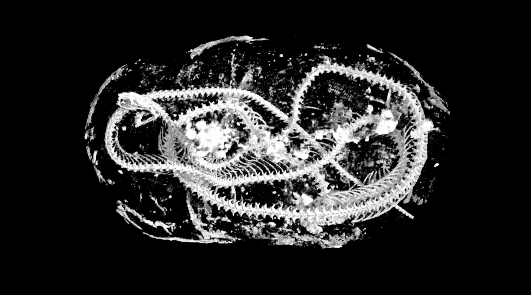
Three mummified animals from ancient Egypt have been digitally unwrapped and dissected by researchers using high-resolution 3D scans that give unprecedented detail about the animals’ lives — and deaths — over 2000 years ago. The three animals — a snake, a bird and a cat — are from the collection held by the Egypt Centre at Swansea University. Previous investigations had identified which animals they were, but very little else was known about what lay inside the mummies. Now, thanks to X-ray micro CT scanning, which generates 3D images with a resolution 100 times greater than a medical CT scan, the animals’ remains can be analyzed in extraordinary detail, right down to their smallest bones and teeth. Credit: Swansea University
Scans give clues to how they lived and died.
Three mummified animals from ancient Egypt have been digitally unwrapped and dissected by researchers, using high-resolution 3D scans that give unprecedented detail about the animals’ lives — and deaths — over 2000 years ago.
The three animals — a snake, a bird and a cat — are from the collection held by the Egypt Centre at Swansea University. Previous investigations had identified which animals they were, but very little else was known about what lay inside the mummies.
Now, thanks to X-ray micro CT scanning, which generates 3D images with a resolution 100 times greater than a medical CT scan, the animals’ remains can be analyzed in extraordinary detail, right down to their smallest bones and teeth.
The team, led by Professor Richard Johnston of Swansea University, included experts from the Egypt Centre and from Cardiff and Leicester universities.
The ancient Egyptians mummified animals as well as humans, including cats, ibis, hawks, snakes, crocodiles, and dogs. Sometimes they were buried with their owner or as a food supply for the afterlife.
But the most common animal mummies were votive offerings, bought by visitors to temples to offer to the gods, to act as a means of communication with them. Animals were bred or captured by keepers and then killed and embalmed by temple priests. It is believed that as many as 70 million animal mummies were created in this way.
Although other methods of scanning ancient artifacts without damaging them are available, they have limitations. Standard X-rays only give 2-dimensional images. Medical CT scans give 3D images, but the resolution is low.
Micro CT, in contrast, gives researchers high-resolution 3D images. Used extensively within materials science to image internal structures on the micro-scale, the method involves building a 3D volume (or ‘tomogram’) from many individual projections or radiographs. The 3D shape can then be 3D printed or placed into virtual reality, allowing further analysis.
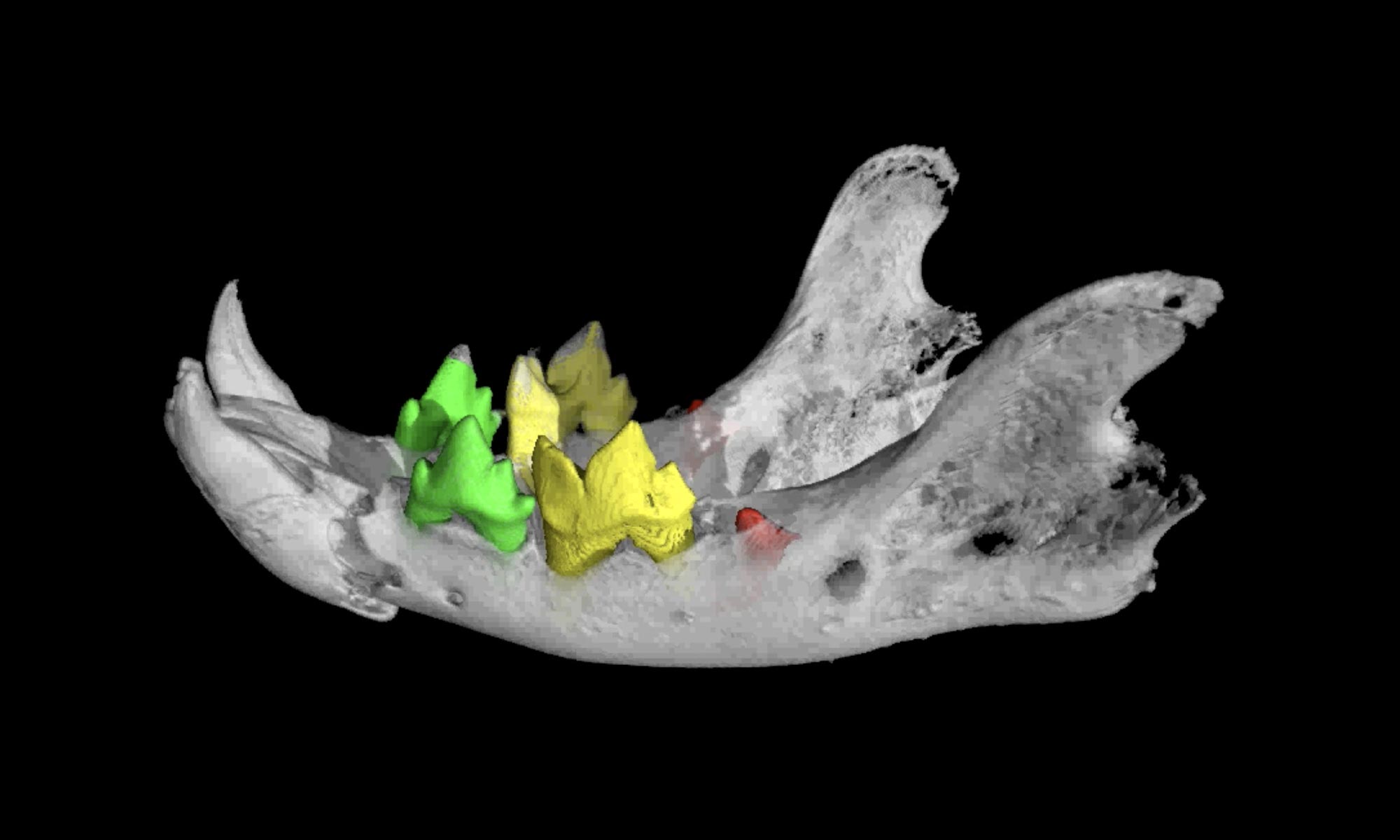
The team, using micro CT equipment at the Advanced Imaging of Materials (AIM) facility, Swansea University College of Engineering, found:
- The cat was a kitten of less than 5 months, according to evidence of unerupted teeth hidden within the jaw bone.
- Separation of vertebrae indicate that it had possibly been strangled
- The bird most closely resembles a Eurasian kestrel; micro CT scanning enables virtual bone measurement, making accurate species identification possible
- The snake was identified as a mummified juvenile Egyptian Cobra (Naja haje).
- Evidence of kidney damage showed it was probably deprived of water during its life, developing a form of gout.
- Analysis of bone fractures shows it was ultimately killed by a whipping action, prior to possibly undergoing an ‘opening of the mouth’ procedure during mummification; if true this demonstrates the first evidence for complex ritualistic behavior applied to a snake.
Professor Richard Johnston of Swansea University College of Engineering, who led the research, said:
“Using micro CT we can effectively carry out a post-mortem on these animals, more than 2000 years after they died in ancient Egypt.
With a resolution up to 100 times higher than a medical CT scan, we were able to piece together new evidence of how they lived and died, revealing the conditions they were kept in, and possible causes of death.
These are the very latest scientific imaging techniques. Our work shows how the hi-tech tools of today can shed new light on the distant past.”
Dr. Carolyn Graves-Brown from the Egypt Centre at Swansea University said:
“This collaboration between engineers, archaeologists, biologists, and Egyptologists shows the value of researchers from different subjects working together.
Our findings have uncovered new insights into animal mummification, religion, and human-animal relationships in ancient Egypt.”
###
Reference: “Evidence of diet, deification, and death within ancient Egyptian mummified animals” by Richard Johnston, Richard Thomas, Rhys Jones, Carolyn Graves-Brown, Wendy Goodridge and Laura North, 20 August 2020, Scientific Reports.
DOI: 10.1038/s41598-020-69726-0
The authors respectfully acknowledge the people of ancient Egypt who created these artifacts.
Never miss a breakthrough: Join the SciTechDaily newsletter.
Follow us on Google and Google News.