
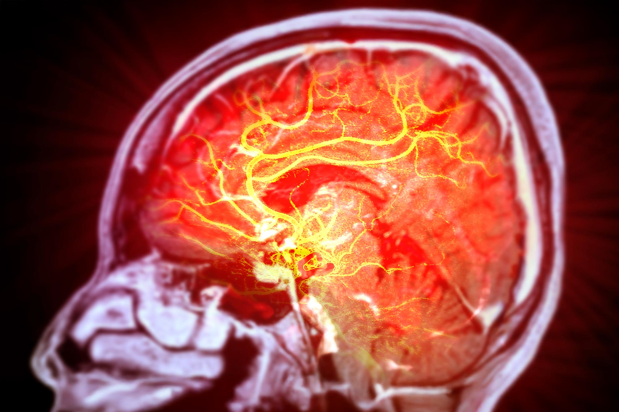
A protein buildup damaged the brain’s protective barrier in animal experiments, pointing to a possible target for Alzheimer’s treatment.
Roughly one in five people carries at least one copy of APOE ε4, the strongest common genetic risk factor for Alzheimer’s disease. Yet many people with the variant remain mentally sharp into their 80s without developing the disease. Understanding what protects some of them could help researchers find ways to protect others.
APOE ε4 carriers accumulate unusually high levels of a protein called fibronectin at the blood-brain barrier, a tightly packed layer of cells around brain blood vessels that helps remove toxic substances. Two years ago, Columbia University researchers discovered that a rare mutation in FN1, the gene that encodes fibronectin, limits this buildup and its damage, helping protect some carriers against Alzheimer’s. A drug that mimics that effect might offer similar protection.
The team’s latest study, published in Nature Aging, found that excess fibronectin can make the barrier leak in animal experiments and traced the damage to disrupted signals between cells.
“This new study takes us from a genetic clue to a disease mechanism,” says Caghan Kizil, the study’s corresponding author and professor of neurological sciences in the Department of Neurology and the Taub Institute for Research on Alzheimer’s Disease and the Aging Brain at Columbia University Vagelos College of Physicians and Surgeons.
“We knew that changes in fibronectin could protect against Alzheimer’s, but we didn’t know why excess fibronectin was harmful in the first place. This study gives us that detailed mechanism—and with it, ideas for how to reproduce that natural protection.”
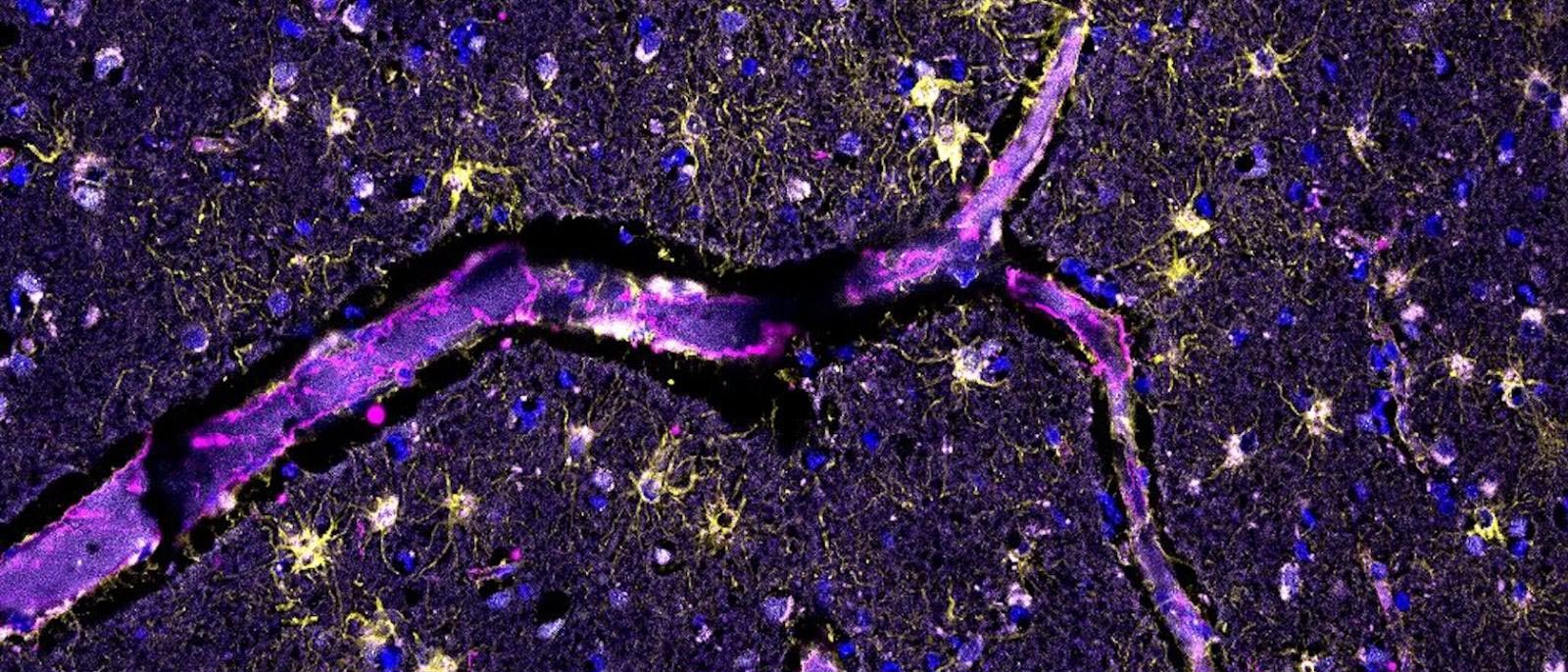
In human brain tissue and cerebrospinal fluid, the liquid surrounding the brain and spinal cord, high fibronectin levels were associated with inflamed astrocytes. These cells, also called astroglia, help maintain the blood-brain barrier. Genetic and epigenetic analyses, which examined genetic variation and gene regulation, also linked FN1 to vascular disease and changes in VEGFA, a growth factor needed to maintain healthy brain blood vessels.
“The convergence of these independent human datasets strengthens the case that fibronectin is part of the APOE ε4-related vascular disease process,” says Badri Vardarajan, associate professor of neurological science in the Department of Neurology, Gertrude H. Sergievsky Center, and Taub Institute, who collaborated on the discovery of the protective FN1 mutation.
Excess fibronectin causes barrier leaks in animals
To investigate how fibronectin affects the barrier, the researchers also used human stem-cell models, three-dimensional vascular cultures, and experiments in zebrafish and mice. Their methods included single-cell sequencing and spatial imaging. Across the systems they examined, the findings pointed to APOE ε4 increasing fibronectin and promoting its accumulation around brain blood vessels.
In mice carrying human APOE ε4, brain fibronectin levels were nearly twice those in mice carrying the normal human APOE version, APOE ε3. The higher levels coincided with evidence that the blood-brain barrier was leaking. That comparison alone could not establish whether fibronectin caused the damage, so the researchers performed a more targeted test. They increased human fibronectin specifically in astrocytes to determine whether excess protein in those cells was enough, by itself, to compromise the barrier.
“The animal experiments allowed us to move beyond association,” says co-first author Prabesh Bhattarai, an associate research scientist in the Kizil lab. “When we increased fibronectin in astroglia, the barrier became permeable. When we reduced fibronectin, barrier function improved.”
The researchers traced the damage to disrupted communication between cells. Excess fibronectin sends abnormal signals through integrins, proteins that sense a cell’s surroundings, and an enzyme called focal adhesion kinase. This disrupts a chain of growth factors called VEGF, HBEGF, and IGF1. These factors allow astrocytes and blood-vessel cells to communicate and work together to maintain the barrier.
“Single-cell analysis allowed us to examine this pathway in the different cells that maintain the brain’s blood vessels,” says co-first author Elanur Yilmaz, an associate research scientist in the Kizil lab. “We observed the same biological pattern across experimental models and human Alzheimer’s tissue, making the mechanism especially compelling.”
The human findings suggest that the same damaging processes likely occur in Alzheimer’s patients who carry APOE ε4, although the evidence that excess fibronectin itself can cause barrier leakage came from the animal experiments.
Targeting fibronectin without disrupting tissue repair
The findings suggest three possible treatment strategies for people with APOE ε4. Researchers could try to prevent excess fibronectin from accumulating, block the harmful signals it triggers, or restore the growth signals needed to keep the blood-brain barrier healthy. The team is exploring these possibilities in laboratory experiments.
Any future treatment would need to target pathological fibronectin while preserving its normal roles in tissue structure and repair. Understanding the mechanism provides a foundation for developing more selective interventions and determining when they might be most useful.
“By showing how fibronectin contributes to early vascular damage, this study gives us new therapeutic targets and potential ways to measure whether treatments are working,” says Richard Mayeux, chair of the Department of Neurology and a collaborator on the project. “Clinically, it connects a major genetic risk factor for Alzheimer’s to an early disease process that may be possible to modify.”
Reference: “Fibronectin mediates APOE4-driven blood–brain barrier dysfunction in Alzheimer’s disease” by Prabesh Bhattarai, Elanur Yilmaz, Elif Öykü Cakir, Huseyin Tayran, Imdadul Haq, Dylan Murphy, Jonah Nadelmann Keller, Quentin Le Grand, Jacqueline O. Eschbach, Hilal Celikkaya, Mehmet I. Cosacak, Hande Yüceer Korkmaz, Annie J. Lee, Verena Haage, Yiyi Ma, Xue Wang, Nastasia Nelson, Weilin Lin, Yixin Zhang, Bengisu Turgutalp, Sherida M. de Leeuw, Ronak Patel, Dörthe Jülich, Özkan İş, Scott A. Holley, Philip L. De Jager, Uwe Freudenberg, Carsten Werner, Peter St George-Hyslop, Nilüfer Ertekin-Taner, Stéphanie Debette, Fanny M. Elahi, Elizabeth S. Fisher, Hemali Phatnani, Andrew F. Teich, Kate Tubbesing, Taylor Bertucci, Sally Temple, Tal Nuriel, Badri N. Vardarajan, Richard Mayeux and Caghan Kizil, 11 September 2026, Nature Aging.
DOI: 10.1038/s43587-026-01204-0
The work was supported by the NIH (grants R01AG067501, RF1AG066107, P30AG066462, and U54AG076040); a Schaefer Research Scholars Award; Taub Institute Grant for Emerging Research; Thompson Family Foundation Program for Accelerated Medicine Exploration in Alzheimer’s Disease and Related Disorders of the Nervous System; American Brain Foundation Cure-One-Cure-Many Award; Carol and Gene Ludwig Family Foundation; Toffler Scholar Program; National Institute on Aging and Department of Veterans Affairs (IK2CX002180); Chan Zuckerberg Initiative; and Rainwater Charitable Foundation.
Never miss a breakthrough: Join the SciTechDaily newsletter.
Follow us on Google and Google News.