A new study reveals that maintaining healthy blood vessels is vital for brain health and could help combat neurodegenerative diseases like Alzheimer’s by enabling earlier detection and potentially guiding new treatments.
Researchers pinpoint regions in the mouse brain susceptible to blood vessel degeneration, shedding light on the link between vasculature and neurodegenerative diseases.
Maintaining healthy blood vessels is important not only for heart health but also for brain health. According to a new study led by Penn State researchers, vascular well-being is crucial in addressing age-related cognitive decline and neurodegenerative disorders such as Alzheimer’s disease. The findings highlight the potentially significant but understudied role that the brain’s vascular network, or energy infrastructure, plays in the onset of neurodegenerative diseases.
They published their work in the journal Nature Communications.
Using advanced imaging techniques, the team developed maps of a mouse brain that illustrate how vascular cells and structures like blood vessels change with age and identified areas that are vulnerable to deterioration. When blood vessels degrade, nerve cells in the brain, called neurons, are starved of energy, causing them to malfunction or die. It can lead to a condition called vascular dementia, the second leading cause of cognitive impairment in older adults, and symptoms like sleep disturbance.
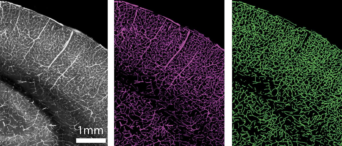
Researchers used advanced imaging techniques to identify vascular cells and structures like blood vessels in mice brains. Credit: Provided by the Kim Lab / Penn State
Early Detection and Understanding of Neurodegenerative Diseases
“With something like Alzheimer’s disease, by the time you can see vascular changes and significant brain shrinkage on an MRI, cell death has already occurred. We need to understand how these cells and structures change before a major catastrophe happens,” said Yongsoo Kim, associate professor of neural and behavioral sciences at Penn State College of Medicine and senior author of the study. “This study provides early signs of neurodegenerative disorders, potentially leading to earlier diagnosis and clues for how we can slow down the aging process and cognitive changes.”
According to Kim, aging is one of the primary factors involved in neurodegenerative disorders.
“Yet, we really don’t have a good baseline understanding of how normal aging itself changes the brain, particularly the brain’s vasculature,” Kim said. And with the aging population in the United States growing, he said it’s critical to understand these changes, especially within the network of blood vessels.
Blood vessels, especially micro-vessels, regulate oxygen and energy supply and waste removal to and from neurons. Despite their importance, Kim said, most existing research focuses on how neuron structure and function degenerate over time, rather than the vasculature. When researchers do study the brain’s vasculature, they’ve primarily examined larger blood vessels or focused on a single, easy-to-access region of the brain, the somatosensory cortex. More importantly, typical neuroimaging techniques, like MRI, don’t provide high enough resolution to see what’s happening in the tiny blood vessels, which make up 80% to 85% of the brain’s vasculature, according to Kim.
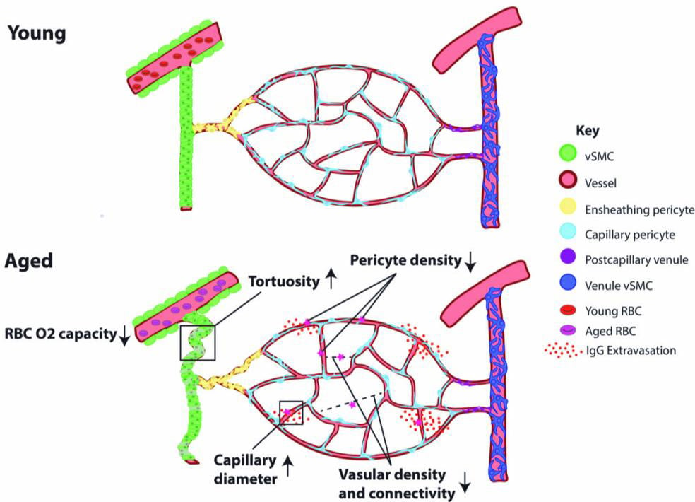
Aged brains show reduced vascular length and branching density, increased radii, reduced pericyte density, leaky blood-brain barrier, and lower oxygen-carrying capacity in the blood compared to young brains. Credit: Provided by the Kim Lab / Penn State
Kim and the research team produced a detailed map of the vascular network of the whole mouse brain using two high-resolution 3D mapping techniques: serial two-photon tomography — a technique that creates a series of stacked 2D images — and light sheet fluorescence microscopy, which images intact 3D samples to visualize the whole brain at a single cell-resolution. They imaged the brains of young and old mice to chart vasculature changes across the brain with normal aging.
“Because we’re doing high-resolution mapping with sufficient resolution, we can reconstruct the whole vascular structure and scan the entire brain to pinpoint areas that undergo selective degeneration with age,” Kim said. “What we found is that the area that most people study showed the least amount of change, whereas profound change happens in areas in the deep areas of the brain. This suggests that we’ve been looking at the wrong area when it comes to aging studies.”
Significant Findings and Future Directions
The images showed that changes in the vascular network don’t occur equally across the brain. Rather, they were concentrated in the basal forebrain, deep cortical layers, and hippocampal network, suggesting these areas are more vulnerable to vascular degeneration. These regions play a role in attention, sleep, memory processing, and storage, among other functions.
As brains age, vascular length and branching density decrease by approximately 10%, indicating that there’s a sparser network to distribute blood. Arteries in older brains also appear more twisted compared to those in younger brains, which can impede blood flow, especially to areas further away from the main arteries like the deep cortical layers, Kim explained.
The team also examined functional changes in vasculature and found that the system responds more slowly in older brains. That means that it can’t provide the neurons with energy as quickly and readily as the cells may need. There’s also a loss of pericytes, a type of cell that regulates blood supply and blood vessel permeability, too. As a result, the blood vessels become “leaky,” compromising the blood-brain barrier.
This study builds on the group’s previous research, where they mapped the vasculature of a young mouse brain. Next, they are studying how Alzheimer’s disease-induced changes in the brain influence vascular health and neuronal function. Ultimately, they said they hope their work will lead to treatments for neurodegenerative disorders.
Reference: “Aging drives cerebrovascular network remodeling and functional changes in the mouse brain” by Hannah C. Bennett, Qingguang Zhang, Yuan-ting Wu, Steffy B. Manjila, Uree Chon, Donghui Shin, Daniel J. Vanselow, Hyun-Jae Pi, Patrick J. Drew and Yongsoo Kim, 30 July 2024, Nature Communications.
DOI: 10.1038/s41467-024-50559-8
Hannah Bennett, dual medical degree and doctoral degree student, and Steffy Manjila, postdoctoral scholar, co-led the study along with Quingguang Zhang, who was assistant research professor at Penn State at the time of the research and is currently assistant professor at Michigan State University, and Yuan-ting Wu, who was previously a research scientist at Penn State and currently project scientist at Cedars-Sinai Medical Center. Other Penn State authors on the paper include: Patrick Drew, professor of engineering science and mechanics, of neurosurgery, of biology and of biomedical engineering and interim director of the Huck Institutes of the Life Sciences; Uree Chon, research technician; Donghui Shin, research technologist; Daniel Vanselow, research project manager; Hyun-Jae Pi, data scientist.
The National Institutes of Health and the American Heart Association funded this work.


Be the first to comment on "Age-Related Cognitive Decline Linked to Blood Vessel Health"