Study is the first to analyze prenatal MRI scans of children later diagnosed with autism.
A new study using prenatal brain scans revealed significant differences in brain structures at around 25 weeks’ gestation between children who were later diagnosed with ASD and those who were not. The study adds to mounting evidence that autism begins in early development and suggests possible opportunities to identify the disorder at an earlier age.
“Earlier detection means better treatment,” said Alpen Ortug, PhD, a postdoctoral research fellow at Athinoula A. Martinos Center for Biomedical Imaging, Massachusetts General Hospital, Harvard Medical School, first author of the study. “Our results suggest that an increased volume of the insular lobe may be a strong prenatal MRI biomarker that could predict the emergence of ASD later in life.”
Ortug will present the research at the American Association for Anatomy annual meeting during the Experimental Biology (EB) 2022 meeting, held in Philadelphia on April 2-5, 2022.
ASD, diagnosed in 1 in 68 children in the U.S., is a complex neurodevelopmental disorder that can cause challenges with communication, cognitive processing, emotional awareness and perception. The causes of ASD are unknown but both genetic and environmental factors are thought to play a role. While early treatment has been shown to improve language and cognitive abilities, current diagnostic tools can only identify the disorder around 18 months of age.
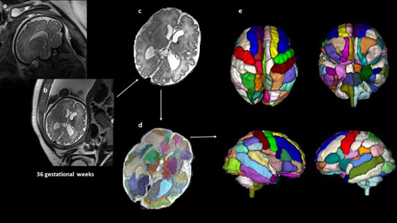
To find out if brain scans taken prenatally could help identify signs of ASD earlier, the researchers retrospectively analyzed 39 fetal MRI brain scans taken at Boston Children’s Hospital. Nine of the children were later diagnosed with ASD, 20 were neurotypical and 10 did not have ASD but had other health conditions that were also observed in the children with ASD. The brain scans had been taken at about 25 weeks’ gestation, on average.
Differences in the Insular Lobe and Other Brain Regions
After preprocessing, the researchers used an atlas-based automated anatomical labeling method to segment the brain scans and then compared the segmented brain regions between the different groups. The biggest differences were found in the brain’s insular lobe, which had a significantly larger volume in the ASD group compared with the other three control groups. The insula is a region deep inside the brain that is thought to have a role in perceptual awareness, social behavior and decision-making, among other functions.
The findings align with other recent studies that have reported changes in the insular cortex in adults with autism and suggests these differences may begin in the womb. The researchers also found that the scans from children with ASD showed a significantly larger amygdala and hippocampal commissure compared with children who had other health conditions but not ASD.
“Given that many genetic and environmental factors could affect the emergence of ASD starting in the fetal stages, it is ideal to identify the earliest signature of brain abnormalities in prospective autism patients,” said Ortug. “To the best of our knowledge, this is the first attempt to semi-automatically segment the brain regions in the prenatal stage in patients who are diagnosed with autism later and compare different groups of controls.”
Ortug conducted the research while in a former position as a postdoctoral research fellow at Boston Children’s Hospital. The study was led by Harvard Medical School Assistant Professor Emi Takahashi, PhD, whose lab recently moved from Boston Children’s Hospital to the Athinoula A. Martinos Center for Biomedical Imaging at Massachusetts General Hospital.
Ortug will present this research from 9:30–9:45 a.m. Tuesday, April 5, in Room 108A, Pennsylvania Convention Center (abstract). Contact the media team for more information or to obtain a free press pass to attend the meeting.
Meeting: Experimental Biology 2022
Never miss a breakthrough: Join the SciTechDaily newsletter.
Follow us on Google and Google News.
2 Comments
Interesting, as long as the “data” is not used to inform the mother to “Abort the baby.” Because the child “MAY” be born with autism. Autism is a blessing, not a curse. More people in the science community, if they don’t already know this, should know this. Autism people think in 5D, which is WHY the ways of the world doesn’t much sense to them. The world is 3D, and non spectrum people are 2D. So, an Autism person becomes best friends with their brain? And actually, can spend all day with their friend the brain, and not care if they don’t engage in human contact.. Spocks’ character was autistic. There is no doubt.
I am proudly Autistic and am a great poet and fanfiction writer. I know 10 different proofs of God and I do not want to be cured, other than to help my sore gut. If Autism is ever eliminated from the Human genepool and/or we start aborting babies with Autism the way we do so manmy, precuous Downs syndrom kids, the world will lose so much! Einstein, Disney, Thomas Jefferson Bill Murry, so many more. Downs Syndrum babies were put here to help us learn to love Autistics were put here to teach us all how to think and everyone in the middle were put here to nourish these gifted people and make use of what we teach.