Researchers have developed a method to study bird brains by creating digital endocasts from empty cranial spaces in bird skeletons.
Using this technique, they discovered that physical brain tissues closely match these digital imprints, allowing detailed studies of brain size and structure across 136 bird species.
Historical Bird Skulls Inform Modern Science
Understanding what birds ‘think’ while they fly is a challenge, but scientists from Australia and Canada are gaining fascinating new insights by studying the structure of their brains.
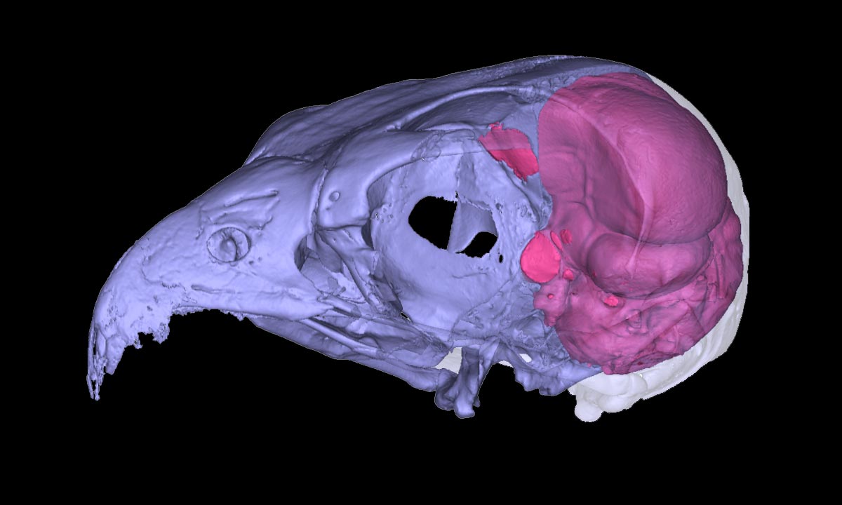
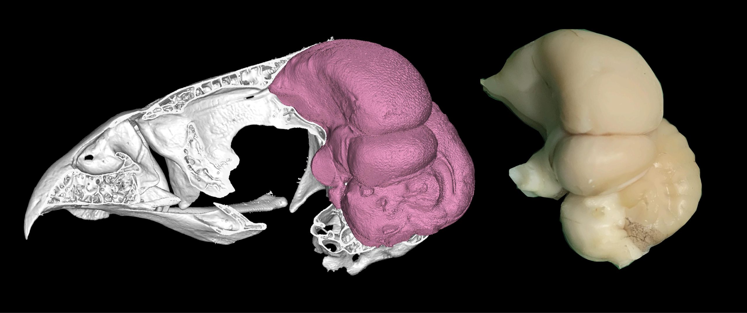
Evolutionary biologists from Flinders University in South Australia and neuroscience researchers from the University of Lethbridge in Canada have joined forces to develop a groundbreaking method for reconstructing the brain structures of both living and extinct birds. They achieve this by creating digital ‘endocasts’—3D models of the space inside a bird’s skull where the brain once resided.
Published in Biology Letters, the study—led by the ‘Bones and Diversity Lab’ at Flinders and the Iwaniuk Lab at the University of Lethbridge—reveals that even the dry skulls of long-dead birds can offer remarkable insights into brain structure. These include details about the size of key brain regions responsible for intelligence and coordination.
This discovery was made by comparing historical microscopic brain sections with digital endocasts, in what is the largest study of its kind, analyzing 136 bird species.
Technological Advances in Bird Brain Research
“This showed that the two correspond so closely that there is no need for the actual brain to estimate a bird’s brain proportions,” says the lead author, Flinders University PhD Aubrey Keirnan.
“While ‘bird brain’ is often used as an insult, the brains of birds are so large that they are practically a braincase with a beak. We decided to test if this also means that the brain’s imprint on the skull reflects the proportions of two crucial parts of the actual brain.”
Joined by researchers at the Department of Neuroscience at the University of Lethbridge in Alberta, Canada, the team scanned the skulls of 136 bird species for which they also had microscopic brain sections or literature data.
This allowed them to determine if the volume of two crucial brain parts, the forebrain, and the cerebellum, corresponds with the surface areas of the endocasts.
The extremely tight match between the ‘real’ and the ‘digital’ brain volumes surprised the researchers.
Timelapse of PhD student Aubrey Keirnan mounting a serially sectioned bird brain onto slides so that they can be measured and analyzed under a microscope at the Iwaniuk lab in Canada. Credit: Aubrey Keirnan
The Future of Neuroanatomy: Digital Insights into Extinct Species
“We used computed microtomography to scan the bird skulls. This allows us to digitally fill the brain cavity to get the brain’s imprint, also called an ‘endocast’,” says senior co-author Associate Professor Vera Weisbecker, from Flinders University’s College of Science and Engineering.
“The correlations are nearly 1:1, which we did not expect. But this is excellent news because it allows us to gather insight into the neuroanatomy of elusive, rare, and even extinct species without ever even seeing their brains.”
Associate Professor Vera Weisbecker says that advanced digital technologies are providing ever-improving access to some of the oldest puzzles in animal diversity.
“The great thing about digital endocasts is that they are non-destructive. In the old days, people needed to pour liquid latex into a brain case, wait for it to set, and then break the skull to get the endocast.
“Using non-destructive scanning not only allows us to create endocasts from the rarest of birds, it also produces digital files of the skulls and endocasts that can be shared with scientists and the public.”

With an extensive background in bird brain research, University of Lethbridge Professor Andrew Iwaniuk, who co-led this study with Associate Professor Weisbecker, says he did not expect such a clear correlation between brain tissue and endocasts.
“While most of the telencephalon (outer part of the forebrain) is visible from the outer surface, a substantial portion of the cerebellum is obscured by this region. Additionally, the avian cerebellum has ‘folds’ which are often obstructed by a large blood vessel called the occipital sinus,” says Professor Iwaniuk.
“Given that the degree of obscurity can vary between species, I did not expect a strong correlation between endocast surface area and brain volume across all species.”
Professor Iwaniuk adds that the study provides support for existing research by other scientists – including for critically endangered modern birds or perhaps even species gone extinct.
However, the team says that it remains to be seen how well the data can be applied to dinosaurs, which are the birds’ closest extinct relatives.
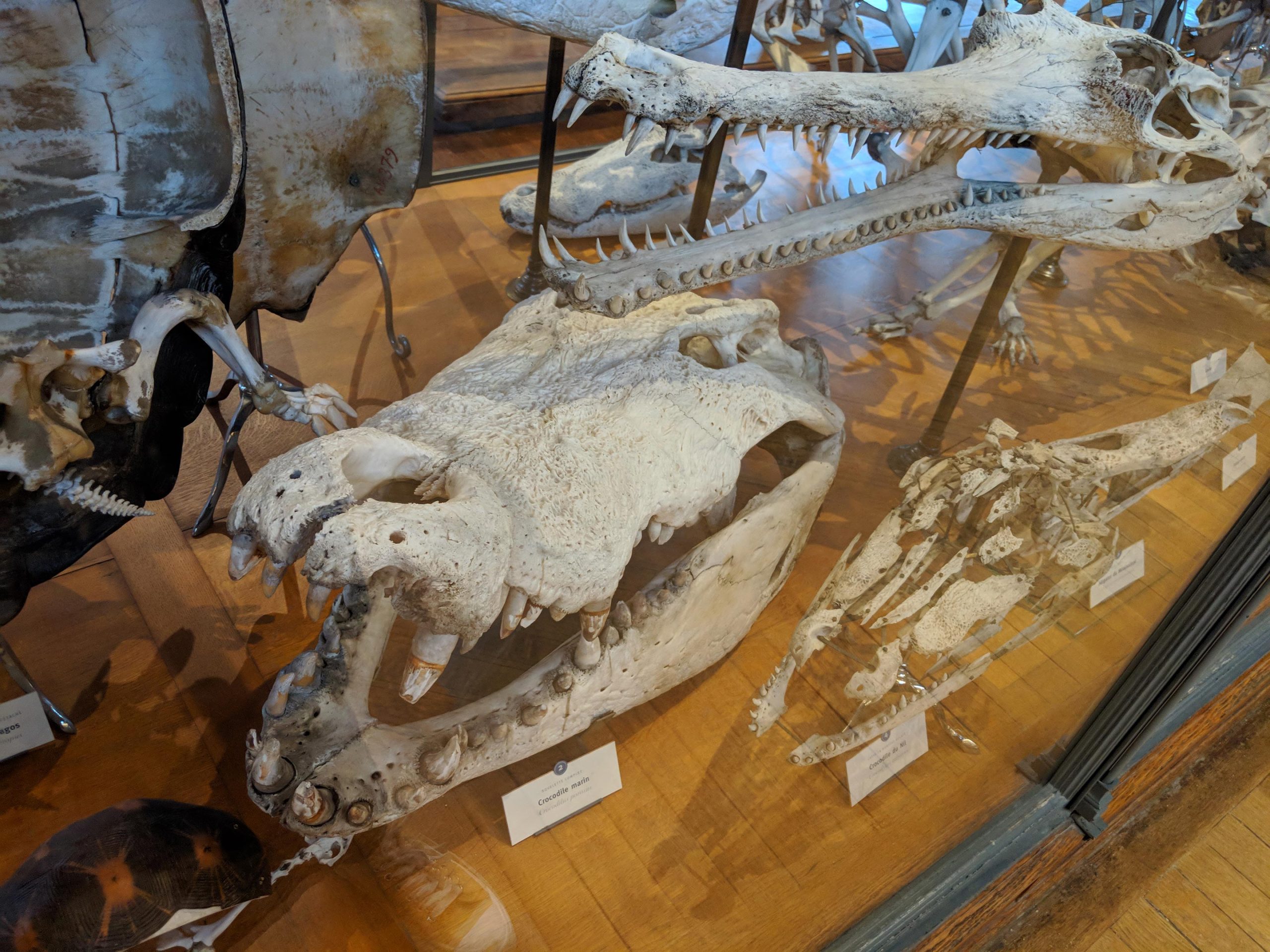
“For example, crocodiles are the closest living relatives of birds, but their brains look nothing like that of a bird – and their brains do not fill the braincase enough to be as informative,” adds Ms. Keirnan.
Reference: “Avian telencephalon and cerebellum volumes can be accurately estimated from digital brain endocasts” by Aubrey R. Keirnan, Felipe Cunha, Sara Citron, Gavin Prideaux, Andrew N. Iwaniuk and Vera Weisbecker, 1 January 2025, Biology Letters.
DOI: 10.1098/rsbl.2024.0596
Never miss a breakthrough: Join the SciTechDaily newsletter.
Follow us on Google and Google News.