Neuroscientists found that mice, like humans, have distinct processing streams in visual cortical areas and plan to use this to explore more deeply the neurological basis of behavior. Harvard researchers are studying how distinct brain areas respond to different visual stimuli in the visual cortex of mice. They believe the findings will open new avenues of brain exploration and will shed light on mammalian vision.

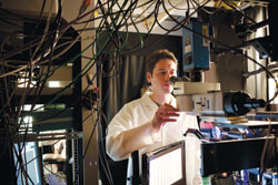
Research Fellow Marc Andermann studies mouse vision in the lab of Neurobiology Professor Clay Reid. Credit: Harvard Medical School
It’s hard to be a mouse. You’re a social animal, but your fellows are small and scattered. You’re a snack to a bestiary of fast, eagle-eyed predators, not least the eagle. You’re fast too, but your spatial vision is poor — around 20/200. So what’s a mouse to do?
For starters, pay attention.
Or specifically, pay attention to small, slow-moving shapes — like other mice — and big, fast ones — the kind you see when you’re running for your life. Evolve specialized regions of the brain to process such cues. And keep moving.
Those regions are the subject of imaging research at Harvard Medical School aimed at revealing how distinct brain areas respond to different visual stimuli in the visual cortex of mice. The findings not only shed new light on mammalian vision, but also open a new avenue for exploring the mysteries of the brain.
“The cortex is really the crowning glory of brain evolution; it’s what allows us to think and be human,” said Clay Reid, HMS professor of neurobiology and senior author on a paper reporting the findings December 22 in the journal Neuron. “And we know that one of the hallmarks of our cortex is that there are dozens of distinct cortical areas that do different things. And the best-studied part of the cortex in any species, is the visual system, particularly the multiple visual areas in primates, including ourselves.”
To explore how the mouse brain responded to different visual stimuli, the researchers tagged neurons in and around the primary visual cortex with a chemical that fluoresced when the neurons fired. They then showed the mice patterns that moved across a screen at different speeds, recording neural activity as the mice watched. The researchers found that the primary visual cortex contained a mix of neurons that responded to various types of stimuli, while adjacent areas were more specialized, some for larger, faster visual cues, others for smaller, slower ones.
The types of specialization made sense, the researchers said, when one considers the challenges mice face, such as identifying other mice, or navigating at high speeds. “You have to think like a mouse to ask what the different mouse visual areas might be doing,” said Mark Andermann, a neurobiology postdoctoral fellow in the Reid laboratory, and lead author on the paper.
The research, among the first to examine how different cortical areas in the mouse are specialized for processing different aspects of visual inputs, draws together two key areas of brain science: On the one hand, vision studies in humans and other primates have propelled the field of cognitive neuroscience over the last half-century. Scientists know today that primates, including humans, have 20 or more different areas that are specialized to perceive such things as color, motion, and complex forms like faces.
At the same time, the mouse is rapidly becoming the best-studied species in neuroscience, including studies of the cortex. So discovering that, like us, mice have distinct processing streams in visual cortical areas lays the foundation for new efforts in cognitive neuroscience that can exploit the powerful genetic tools available only in the mouse. As a result, researchers can explore more deeply the neurological basis of behavior.
The mouse brain has other features that make it attractive to researchers. Where the human brain resembles a giant, wrinkly walnut, the peanut-sized mouse brain is also peanut smooth, the better for imaging. And where the human brain has about 100 billion neurons with about 5 billion in visual areas, the mouse has only 100 million neurons, with a less daunting figure of 1 million to 2 million in visual areas.
“Even though the visual cortex is more than 1,000 times smaller in mice than in humans,” Reid said, “it’s amazing that we have so many things in common.”
Reference: “Functional Specialization of Mouse Higher Visual Cortical Areas” by Mark L. Andermann, Aaron M. Kerlin, Demetris K. Roumis, Lindsey L. Glickfeld and R. Clay Reid, 22 December 2011, Neuron.
DOI: 10.1016/j.neuron.2011.11.013


Be the first to comment on "Neuroscientists Study Cortical Areas Specialized in Processing Visual Inputs in Mice"