
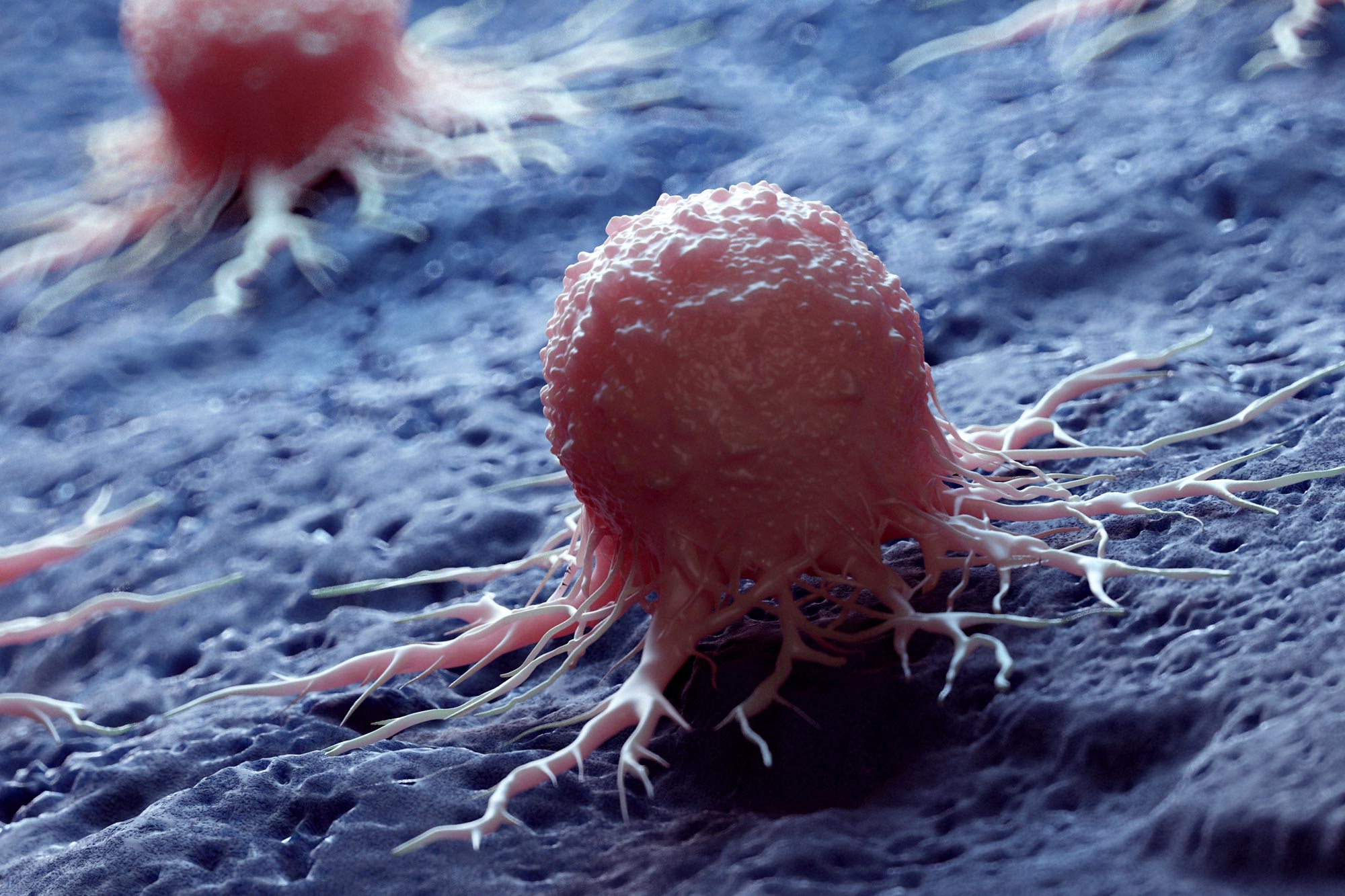
Dormant breast cancer cells may survive treatment inside protective cellular neighborhoods that require different therapeutic strategies.
Inside a breast tumor, rapidly multiplying cancer cells can sit beside others that appear almost inactive. Researchers from the MRC Laboratory of Medical Sciences (LMS), Imperial College London and UCL Genetics Institute have now mapped these contrasting cellular regions in detail, identifying separate neighborhoods of proliferating and dormant cells.
Published in Genome Medicine, the study found that immune and connective tissue cells often surround the dormant cancer cells, potentially creating a protective environment that helps them withstand treatment. The results suggest that preventing tumor growth and later recurrence may require therapies that address both cancer cell states and the local conditions that support their survival.
A tumor contains far more than one uniform mass of cancer cells. It also includes immune cells, newly-formed blood vessels, and quiescent cancer cells that have temporarily stopped dividing. These dormant cells can avoid treatments aimed at active growth, then later awaken and contribute to cancer spreading or returning.
Researchers at the LMS, Imperial and UCL used publicly available information to investigate where quiescent cells are located in untreated tumors, what distinguishes them and which cells occupy the surrounding space. Their map revealed defined pockets of dormant cells enclosed by neighboring cells that may serve as a protective guard.
“Quiescent cancer cells are very dangerous,” explains Dr. Alexis Barr, co-lead author and head of the Cell Cycle Control group at the LMS. “These cells can hide from chemotherapy and then remain in this dormant quiescent state in the tumor, and then later reactivate to drive proliferation.”
Some cancer cells enter quiescence when the demands of rapid tumor growth outpace supplies of nutrients and blood. By pausing their activity during these stressful conditions, they may survive until the environment becomes more favorable, potentially after treatment has ended. Alexis adds, “If we want to achieve long-term control of peoples’ tumors and prevent tumor relapse, we have to focus on these dormant quiescent cancer cells, and have to understand more about them.”
Mapping exposes resistant cells before treatment
Alexis collaborated with Dr. Maria Secrier’s computational biology group at UCL to chart the tumor together with its immune and support cells. The researchers combined single-cell RNA sequencing, which revealed which genes each cell was using, with spatial transcriptomics, which showed where those cells and their neighbors were positioned inside the tumor.
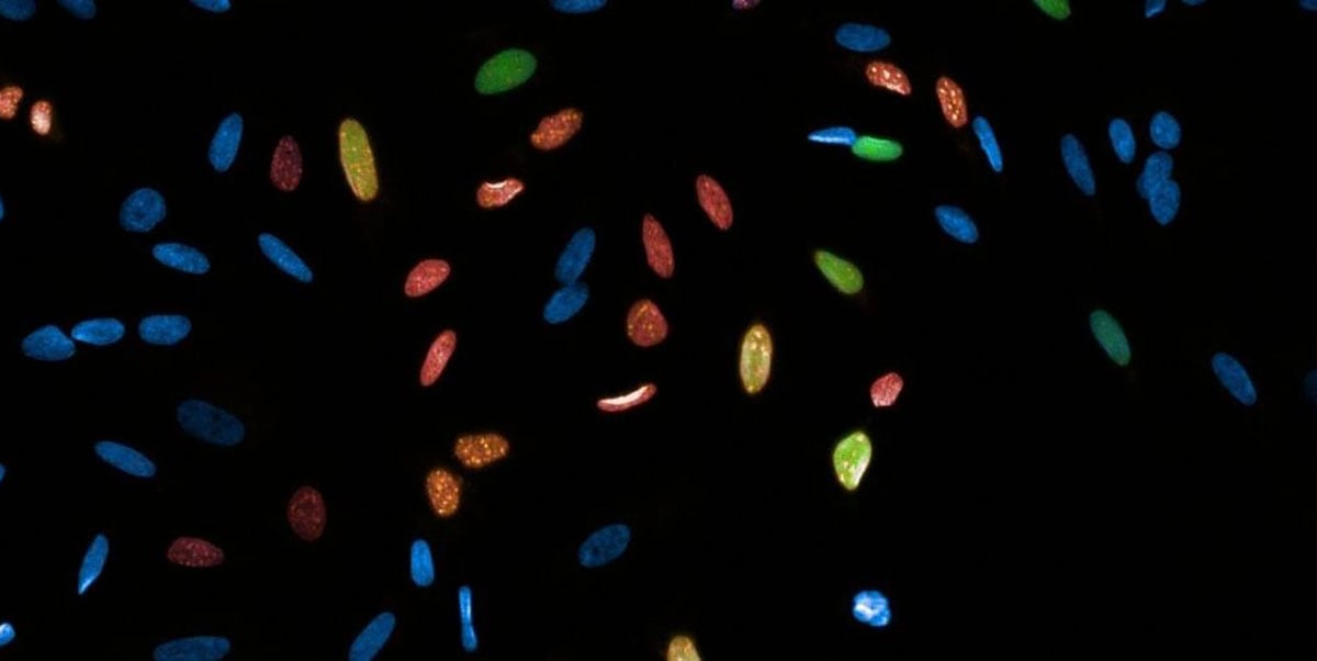
“We found cells that resemble therapy-resistant cells already residing in the tumor before we give any treatment,” says Maria, indicating that characteristics associated with resistance may already exist within a tumor rather than emerging only after therapy begins. These cells appeared in aggressive tumors as well as more slowly developing forms, which was unexpected because quiescence had previously been linked mainly to slower-growing disease.
Support cells may shield dormant cancer
The analysis also examined the many support cells recruited into the tumor environment. A consistent pattern emerged: dormant cancer cells were frequently positioned beside CXCL10-positive macrophages (a type of immune cell) and myofibroblastic cancer-associated fibroblasts (a type of tumor-supporting cell).
These neighboring cells may have been recruited to protect the dormant population, possibly forming a barrier that limits access by cancer-killing immune cells or treatments.
“The cancer cells are really encapsulated within these areas of macrophages and fibroblasts that we think act as shields for these dormant cancer cells,” said Maria. “But we don’t yet know the direction of cause and effect: whether the surrounding cells push cancer cells into dormancy or if the cancer cells attract or alter their surroundings. It’s very likely coming from both sides.”
Different regions may need different drugs
Many chemotherapy drugs work best against cells that are dividing quickly, leaving quiescent cells less exposed to their effects. The findings indicate that active and dormant tumor regions may therefore respond differently to treatment.
Within the dormant niches, the researchers detected greater activity in the complement pathway (a part of the immune system). That pattern may create an opportunity for therapies directed at this pathway. The macrophages and fibroblasts surrounding the cancer cells could provide another possible target, although experiments are still needed to determine whether they actively maintain dormancy.
“Different parts of the tumor will likely respond to different drugs,” said Maria. “If we understand what drug combinations we can use to target both the proliferative and the dormant areas, potentially that could be more successful than current therapies. This is giving us a first insight into how we can then intervene with different therapeutics that specifically target different areas of the tumor where the cells have adapted and have evolved differently.”
Alexis said, “It is clearly important to focus on proliferative cancer cells, but we also need to understand this population of quiescent dormant cancer cells. And that’s been less studied.”
The study’s predictions must now be tested experimentally. Still, locating treatment-resistant regions that exist before therapy and identifying the cells that support them could eventually guide drug combinations designed to attack both active and dormant parts of a tumor. Mapping quiescent cells may be an important step toward cancer treatments with more lasting effects.
Reference: “Spatial ecology of breast cancer reveals co-evolution of proliferative and dormant niches” by Cenk Celik, William A. Weston, Thais de Moraes-Lacerda, Eloise Withnell, Shi Pan, Tooki Chu, John Labbadia, Alexis R. Barr and Maria Secrier, 9 July 2026, Genome Medicine.
DOI: 10.1186/s13073-026-01711-0
This work was primarily funded by a UKRI Future Leaders Fellowship, the Medical Research Council and the Biotechnology and Biological Sciences Research Council.
Never miss a breakthrough: Join the SciTechDaily newsletter.
Follow us on Google and Google News.
1 Comment
“Comparing three recent breakthrough discoveries—on arginine-driven MHC-I flag presentation (Cell), the glycocalyx ‘sugar shield’ (Science Advances), and dormant cell ‘cellular neighborhoods’ (Genome Medicine)—exposes how academic nomenclature silos can obscure the bigger picture. Different labs are describing three distinct tiers of an integrated defense strategy without recognizing they are looking at the same system:
Tier 1 (Internal Signaling): The Cell study demonstrates how arginine fuels internal MHC-I antigen assembly—showing how metabolic starvation suppresses the recognition signal from within.
Tier 2 (Membrane Surface Barriers): The Science Advances study reveals how a dense glycocalyx sugar coating creates a physical and electrostatic barrier, deflecting T cells even when surface antigens are present.
Tier 3 (Tissue-Level Microenvironments): The Genome Medicine study maps how dormant cancer cells co-opt surrounding fibroblasts and macrophages to build protective ‘gated communities’ that block drug delivery and immune infiltration at the tissue scale.
The Takeaway: Cell biology, immunology, and oncology are observing a unified defense-in-depth architecture—internal signal suppression, membrane-level physical shielding, and spatial neighborhood isolation.
Real therapeutic breakthroughs will happen when we stop treating these as separate biochemical anomalies and start using cross-disciplinary tools to lower boundary impedance at the tissue, membrane, and metabolic levels simultaneously.”