Caltech’s improved photoacoustic imaging technology, PACTER, simplifies procedures, enables 3D imaging, and reduces operational complexity, marking a significant advancement in medical imaging.
There are times when scientific progress comes in the form of discovering something completely new. Other times, progress boils down to doing something better, faster, or more easily.
New research from the lab of Caltech’s Lihong Wang, the Bren Professor of Medical Engineering and Electrical Engineering, is the latter. In a paper published in the journal Nature Biomedical Engineering, Wang and postdoctoral scholar Yide Zhang show how they have simplified and improved an imaging technique they first announced in 2020.
That technique, a form of photoacoustic imaging technology called PATER (Photoacoustic Topography Through an Ergodic Relay), is a specialty of Wang’s group.
Improvements in Photoacoustic Imaging
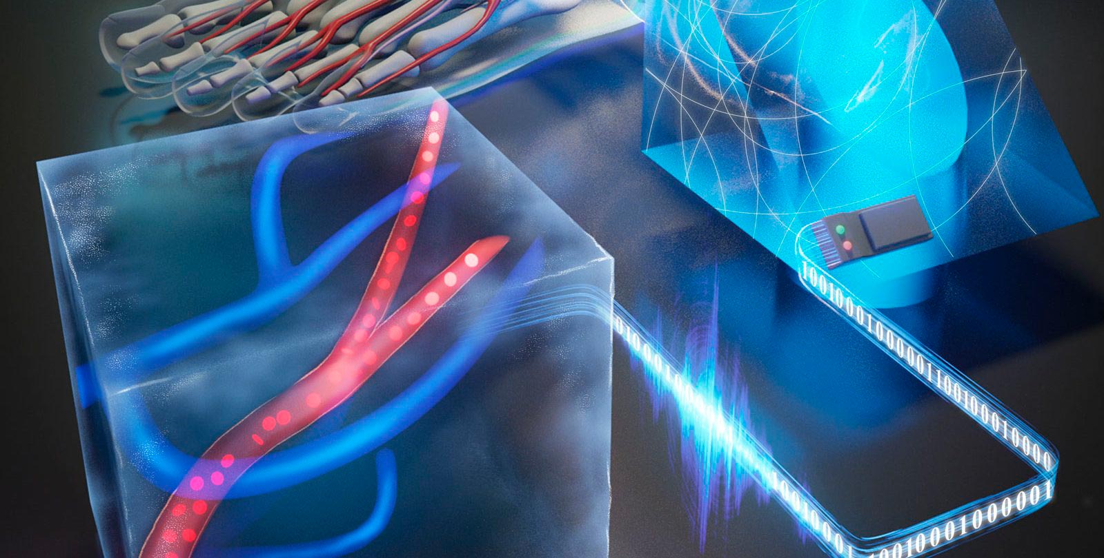
In photoacoustic imaging, laser light is pulsed into tissue where it is absorbed by the tissue’s molecules, causing them to vibrate. Each vibrating molecule serves as a source of ultrasonic waves that can be used to image the internal structures in a fashion similar to how ultrasound imaging is performed.
However, photoacoustic imaging is technologically challenging because it produces all its imaging information in one short burst. To capture that information, early versions of Wang’s photoacoustic imaging technology required arrays of hundreds of sensors (transducers) to be pressed against the surface of the tissue being imaged, which made the technology complicated and expensive.
Wang and Zhang reduced the number of required transducers by using a device called an ergodic relay, which slows down the rate at which information (in the form of vibrations) flows into a transducer. As explained in a previous story about PATER:
In computing, there are two main ways to transmit data: serial and parallel. In serial transmission, the data are sent in a single stream through one communication channel. In parallel transmission, several pieces of data are sent at the same time using multiple communication channels.
The two types of communication are roughly analogous to the way cash registers might be used in a store. Serial communication would be like having one cash register. Everyone gets in the same line and sees the same cashier. Parallel communication would be like having several registers and a line for each.
The system Wang designed with 512 sensors is similar to the store with many cash registers. All of the sensors are working at the same time, with each taking in part of the data about the ultrasonic vibrations generated by the laser pulse.
Since the ultrasonic vibrations from the system come in one short burst, a single sensor would be overwhelmed if it were used to try and collect all the data in that short amount of time. That’s where the ergodic relay comes in.
As Wang describes it, an ergodic relay is a sort of chamber around which sound can echo. When the ultrasonic vibrations pass through the ergodic relay, they are stretched out in time. To return to the cash-register metaphor, it would be like having another employee assisting the single cashier by telling the customers to walk a few laps around the store until the cashier is ready to see them, so the cashier does not become overwhelmed.
PACTER: The Next Evolution
The latest version of this technology, called PACTER (Photoacoustic Computed Tomography Through an Ergodic Relay) goes even further, allowing the system to operate using a single transducer that, through the use of software, can collect as much data as 6,400 transducers.
PACTER improves on PATER in two other ways, says Wang, who is also the Andrew and Peggy Cherng Medical Engineering Leadership Chair and executive officer for medical engineering.
One improvement is that PACTER can create three-dimensional images, whereas PATER can only generate 2D images. This was enabled by the development of improved software.
“Transitioning to 3D imaging significantly escalates the data requirement. The challenge was funneling the immensely increased data through a single transducer,” Zhang says. “Our solution emerged by altering our approach. Rather than a direct and computationally intensive method of reconstructing 3-D images from the single-transducer data, we first expanded one transducer into thousands of virtual ones. This idea simplified the process of 3D image reconstruction, aligning it more closely with the traditional methods in our photoacoustic imaging.”
Secondly, unlike PATER, PACTER does not need to be calibrated each time it is used.
“With PATER, we had to calibrate it each time to use it and that’s just not practical. We got rid of this per-use single-time calibration,” Wang says.
Calibration was needed because when the system fires a pulse of laser light into tissue, an “echo” of that pulse would bounce back into the transducer, preventing it from sensing direct ultrasound information.
Wang says PACTER gets around that issue by adding something called a delay line to the system. The delay line forces the echo to take a longer physical path on its way back to the transducer so that it arrives after the direct ultrasound information has been received.
“Even though I always said this was possible, I knew it would be challenging,” Wang says.
The paper describing the work, “Ultrafast longitudinal imaging of haemodynamics via single-shot volumetric photoacoustic tomography with a single-element detector,” appears in the November 30 issue of Nature Biomedical Engineering. Co-authors are Peng Hu (PhD ’23), former graduate student in medical engineering; Lei Li (PhD ’19), former postdoc in medical engineering; Rui Cao, postdoc in medical engineering; Anjul Khadria, former postdoc in medical engineering; Konstantin Maslov, former staff scientist at Caltech; Xin Tong, graduate student in medical engineering; and Yushun Zeng, Laiming Jiang, and Qifa Zhou of USC.
Funding for the research was provided by National Institutes of Health.
Never miss a breakthrough: Join the SciTechDaily newsletter.
Follow us on Google and Google News.