
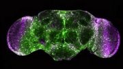
Brain tissue
These findings may have implications for brain disease, disorders.
Scientists at the Krembil Brain Institute, part of University Health Network (UHN), in collaboration with colleagues at the Centre for Addiction and Mental Health (CAMH), have used precious and rare access to live human cortical tissue to identify functionally important features that make human neurons unique.
This experimental work is among the first of its kind on live human neurons and one of the largest studies of the diversity of human cortical pyramidal cells to date.
“The goal of this study was to understand what makes human brain cells ‘human,’ and how human neuron circuitry functions as it does,” says Dr. Taufik Valiante, neurosurgeon, scientist at the Krembil Brain Institute at UHN and co-senior author on the paper.
“In our study, we wanted to understand how human pyramidal cells, the major class of neurons in the neocortex, differ between the upper and bottom layers of the neocortex,” says Dr. Shreejoy Tripathy, a scientist with the Krembil Centre for Neuroinformatics at CAMH and co-senior author on this study.
“In particular, we wanted to understand how electrical features of these neurons might support different aspects of cross-layer communication and the generation of brain rhythms, which are known to be disrupted in brain diseases like epilepsy.”
With consent, the team used brain tissue immediately after it had been removed during routine surgery from the brains of patients with epilepsy and tumors. Using state-of-the-art techniques, the team was then able to characterize properties of individual cells within slices of this tissue, including visualizations of their detailed morphologies.
“Little is known about the shapes and electrical properties of living adult human neurons because of the rarity of obtaining living human brain tissue, as there are few opportunities other than epilepsy surgery to obtain such recordings,” says Dr. Valiante.
To keep the resected tissue alive, it is immediately transferred into the modified cerebrospinal fluid in the operating room then taken directly into the laboratory where it is prepared for experimental characterization.
It is rare to study human tissue because accessing human tissue for scientific inquiries requires a tight-knit multidisciplinary community, including patients willing to participate in the studies, ethicists ensuring patient rights and safety, neurosurgeons collecting and delivering samples, and neuroscientists with necessary research facilities to study these tissues.
After initial analysis, members of the Krembil Centre for Neuroinformatics used further large-scale data analysis to identify the properties that distinguished neurons in this cohort from each other. These properties were then compared to those from other centers doing similar work with human brain tissue samples, including the Allen Institute for Brain Sciences in Seattle, Washington.
Noted in the team’s findings:
- A massive amount of diversity among human neocortical pyramidal cells
- Distinct electrophysiological features between neurons located at different layers in the human neocortex
- Specific features of deeper-layer neurons enabling them to support aspects of across-layer communication and the generation of functionally important brain rhythms
The teams also found notable and unexpected differences between their findings and similar experiments in pre-clinical models, which Dr. Tripathy believes is likely reflective of the massive expansion of the human neocortex over mammalian and primate evolution.
“These results showcase the notable diversity of human cortical pyramidal neurons, differences between similarly classified human and pre-clinical neurons, and a plausible hypothesis for the generation of human cortical theta rhythms driven by deep layer neurons,” says Dr. Homeira Moradi Chameh, a scientific associate in Dr. Valiante’s laboratory at Krembil Brain Institute and lead author on the study.
In total, the team was able to characterize over 200 neurons from 61 patients, reflecting the largest dataset of its kind to date and encapsulating almost a decade’s worth of painstaking work at UHN and the Krembil Brain Institute.
“This unique data set will allow us to build computational models of the distinctly human brain, which will be invaluable for the study of distinctly human neuropathologies,” says Dr. Scott Rich, a postdoctoral research fellow in Dr. Valiante’s laboratory at the Krembil Brain Institute and co-author on this work.
“For instance, the cellular properties driving many of the unique features identified in these neurons are known to be altered in certain types of epilepsy. By implementing these features in computational models, we can study how these alterations affect dynamics at the various spatial scales of the human brain related to epilepsy, and facilitate the translation of these ‘basic science’ findings back to the clinic and potentially into motivations for new avenues in epilepsy research.”
“This effort was only possible because of the very large and active epilepsy program at the Krembil Brain Institute at UHN, one of the largest programs of its kind in the world and the largest program of its kind in Canada,” says Dr. Valiante.
Reference: “Diversity amongst human cortical pyramidal neurons revealed via their sag currents and frequency preferences” by Homeira Moradi Chameh, Scott Rich, Lihua Wang, Fu-Der Chen, Liang Zhang, Peter L. Carlen, Shreejoy J. Tripathy and Taufik A. Valiante, 3 May 2021, Nature Communications.
DOI: 10.1038/s41467-021-22741-9


Brain disease like Epilepsy can be cured.This is plesant to have new findings on brain cells neurons.Utilization of remaining fraction of brain cell and stem cell genetation within brain are some facts of worth.Efficient Electro-Therapy is important.Genetic factors contribute a fraction to the diseade can also be healed specificly which are delinked.
New brain cells or neurons have been foud,is a very good news.Cure of disease like epilepsy is now qùite possiblo.Electro-therapy is useful in such disease.Often delinked genetic stuff in brain which makes disease compĺicate must be paid attention.Another fact is reutilise remaing part of brain cell and generatig stem cell to possibilities.
But what happens when you keep a part of someone’s brain alive, is it still a thinking conscious part or is it no longer human?
Im living w/ Aspergers syndrome. Where is that ” disorder” even though negro Jesus made me this way??
This is trash science.
Taking damaged/malfunctioning tissue and pretending they can determine how normal Grey matter works is laughable. If it was like normal tissue these patients wouldn’t be suffering.
The schools where the PI work and got their should be defunded.
Just my 2 cents… Jb
This is trash science.
Taking damaged/malfunctioning tissue and pretending they can determine how normal Grey matter works is laughable. If it was like normal tissue these patients wouldn’t be suffering.
The schools where the PI work and got their should be defunded.
Just my 2 cents… Jb
Scitech daily won’t let me post this comment. I will post the screen shots to every other platform I can.
My goodness. A live human brain? What if unknowingly they were working while their consciousness was still in play. Couldn’t the brain suffer feelings and thoughts of infinite proportion. Say the perception of time was not in tact and they were forced to re-live their whole life or something like that?
As an epileptic, this study is amazing.
I am literally crying, because this makes me so happy and hopeful for the future of all epileptic patients.
This is incredibly unethical; reminding me of Nazi experiments, as well as experiments done in South America 50 years later.
Of course someone with OUT Epilepsy would think this is unethical. How can science grow and cures for diseases be found if they don’t study the most intricate part of the human body? Only idiotic people would think this is unethical. 🤦♀️
When others are willing to participate and consent is given under full truthful disclosure to a humane research need and any gained understanding of knownlegde is at best intentions of its findings documented with its delibertacy to benefit others is not at any point a Frankenstein experiment. With the complexity of the human brain and all the parts of its components ins every changing adapting and working abilities its the highest priority in human well being both mental and physical and surprisingly spiritual to achieve the pinnacle of quality of life. Keep this in mind before you give your irresponsible , arrogant, short sighted opinions. It is the bravest act of any human being to say yes . Because with it there are unknown , adverse, and terrifing consequences.
If someone donates there body to science to help humanity what a great person to give to others and not be stuck on a old ideology or others fears or believes,thats should be respected not used as to discrete people working to improve and advance peoples lives and health go and pray for some compaction and understanding
Giving your body to science is the most selfless thing you can do. Trying to stop others from progressing science and preventing use of their bodies as they like is the most selfish thing anyone can do.
Period.
I’d like to give my thanks to the brave human being who volunteered to this. Your patronage will help us all go farther, thank you.
Hello, I have a Complex disease and I have reached out to research universities about studying my case. I want to give opportunity to understand and observe complex system damage.