
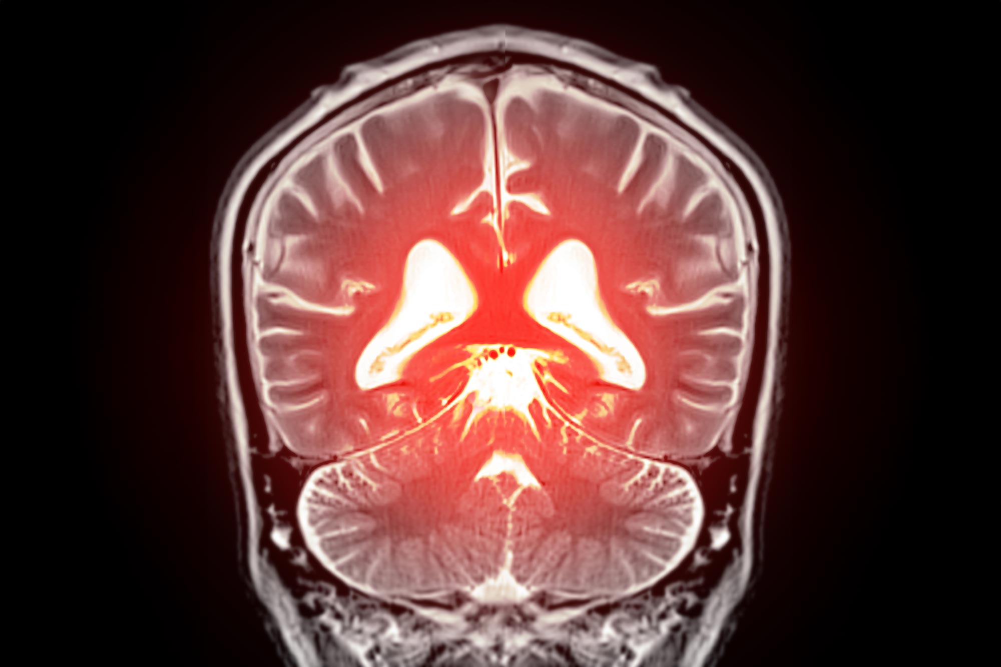
Research involving older adults in India suggests that healthier short-range brain connections may help preserve thinking abilities as gray matter declines.
Two older adults can have a similar amount of gray matter loss yet experience different effects on thinking and memory. Research from the Mark and Mary Stevens Neuroimaging and Informatics Institute (Stevens INI) at the Keck School of Medicine of USC suggests that part of the difference may lie in the condition of superficial white matter, the brain’s short-range wiring that connects nearby regions.
The study, published in Alzheimer’s & Dementia: The Journal of the Alzheimer’s Association, examined brain scans and cognitive test results from 459 community-dwelling adults age 60 and older in India. It is among the first investigations of superficial white matter in a community-based population from a low- and middle-income country.
Superficial white matter forms a thin layer of nerve fibers directly beneath the brain’s outer gray matter. Its short, curved fibers carry signals between neighboring areas of the cerebral cortex, while gray matter contains many of the brain cells responsible for processing information.
“Gray matter and superficial white matter are physically close and may play different roles: gray matter processes information, while superficial white matter helps nearby brain regions communicate,” said Yingxu Liu, PhD, a postdoctoral scholar at the Stevens INI and first author of the study. “Our findings suggest that cognitive health depends not only on how much gray matter is preserved, but also on the condition of the wiring that connects it.”
Healthier brain wiring tracked stronger language skills
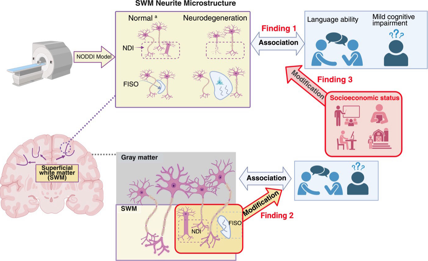
To examine this local wiring in detail, the researchers used an advanced form of diffusion MRI that measures the movement of water through brain tissue. They assessed features including neurite density, which reflects the small projections nerve cells use to send and receive signals, and the amount of freely moving water surrounding them. Reduced neurite density or increased free water can signal tissue disruption associated with myelin loss, inflammation, or swelling.
Participants also took tests measuring language, memory, executive function, and visuospatial ability. Healthier superficial white matter showed its most consistent association with stronger language performance, particularly in frontotemporal regions involved in word recognition, fluent speech, and holding language information in mind.
This animation shows the relationship between superficial white matter and language ability in older Indian adults, with brighter colors indicating stronger associations. Credit: Stevens INI
Healthier wiring may buffer gray matter loss
Measurements of gray matter atrophy were still the strongest overall predictors of cognitive performance. However, the association between gray matter loss and cognition varied with the condition of superficial white matter. When this local wiring had poorer integrity, gray matter loss was more strongly associated with worse language performance and cognitive impairment. When the wiring was healthier, those relationships were weaker.
“The findings point to superficial white matter as a possible source of resilience,” said Leon Aksman, PhD, assistant professor of research neurology at the Stevens INI and senior author of the study. “Two people with a similar degree of gray matter loss may not experience the same cognitive effects if the local connections surrounding that gray matter differ in health. Following participants over time will be essential to test whether preserving these connections can help maintain cognition.”
The link was stronger in some groups
The researchers drew their data from the Harmonized Diagnostic Assessment of Dementia for the Longitudinal Aging Study in India, or LASI-DAD. More than half of the larger LASI-DAD sample has low literacy, and about 60% live in rural communities, populations that have rarely been included in neuroimaging studies.
In this analysis, the association between superficial white matter and language was stronger among people who could not read or read incorrectly, had received no formal education, or lived in rural areas. The findings do not establish that these social conditions caused changes in the brain. Instead, they suggest that lifelong social, educational, health, and environmental experiences may intersect with the aging process in the brain.
One snapshot cannot establish what comes first
Because the researchers assessed participants at a single point in time, they cannot determine whether superficial white matter changes occur before gray matter loss or cognitive decline. Future long-term research will track how the two tissues change together and investigate contributions from vascular health, inflammation, Alzheimer’s-related proteins, and other factors.
“A fuller understanding of brain aging requires research that reflects the world’s social, cultural, and geographic diversity,” said Arthur W. Toga, PhD, director of the Stevens INI and Provost Professor at USC. “By studying an underrepresented population and looking beyond gray matter alone, this work brings us closer to identifying the biological and social factors that may protect cognition across the lifespan.”
Reference: “Superficial white matter and gray matter jointly support cognition among older adults in India” by Yingxu Liu, Kirsten M. Lynch, Miguel Arce Rentería, Emma Nichols, Alden L. Gross, Lindsay C Kobayashi, Neda Jahanshad, John P. John, Harshita V. Vishwakarma, Pranali Khobragade, Joyita Banerjee, Niranjan Khandelwal, Jyoti Dangwal, Sudhir Saxena, Nirod Medhi, Soumik Das, Prudhvinath Reddy, Pratyaksha Rana, Arjun Narula, Saravanan Kannan, Dinesh Patel, A. B. Dey, Sharmistha Dey, Jinkook Lee and Leon M. Aksman, 27 July 2026, Alzheimer’s & Dementia.
DOI: 10.1002/alz.71697
The research was supported by the National Institute on Aging (R01AG080473, RF1AG087965, RF1AG088003, R01AG087513), the National Institute of Mental Health (R01MH134004), the National Institute of Neurological Disorders and Stroke (RF1NS136995), and the Office of the Director of the National Institutes of Health (S10OD032285).
Never miss a breakthrough: Join the SciTechDaily newsletter.
Follow us on Google and Google News.