Scientists rebuilt human brain circuits in the lab and discovered that the thalamus acts as a central organizer of cortical wiring. The findings offer new insight into how brain networks form and why they sometimes go awry.
A team of researchers in Japan has recreated key human neural circuits in a laboratory setting by using tiny, multi-region brain models known as assembloids. These structures are grown from induced pluripotent stem (iPS) cells and combine multiple brain-like regions into a single system. Using this approach, the researchers showed that the thalamus plays a vital role in shaping specialized neural circuits within the human cerebral cortex.
The study was published in the journal Proceedings of the National Academy of Sciences of the United States of America.
Why Neural Circuits in the Cortex Matter
The cerebral cortex is home to many different types of neurons that must communicate smoothly with one another and with other areas of the brain. These interactions support essential mental abilities, including perception, learning, and cognition.
In people with neurodevelopmental conditions such as autism spectrum disorder (ASD), these cortical circuits often develop differently or function improperly. Gaining a clearer understanding of how these neural networks form is an important step toward identifying the biological roots of such disorders and creating more effective treatments.
The Thalamus and Brain Circuit Development
Research in rodents has long suggested that the thalamus plays a key role in organizing cortical neural circuits. What has remained uncertain is how the thalamus and cortex work together to shape these circuits in the human brain.
Directly studying this process in humans presents major ethical and technical challenges, since collecting brain tissue is highly limited. To work around these barriers, scientists have turned to organoids, which are three-dimensional structures grown from stem cells that resemble real organs.
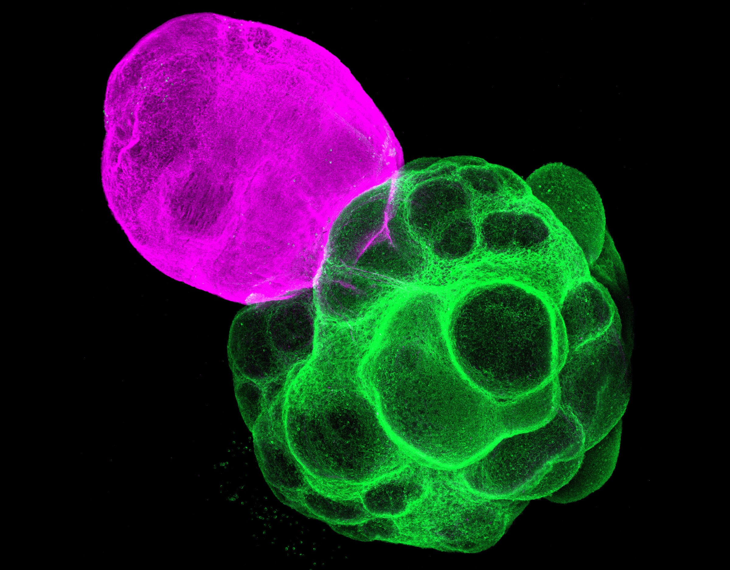
Assembloids Bring Brain Regions Together
While organoids are useful models, a single organoid cannot capture the interactions between multiple brain regions. To address this limitation, researchers use assembloids, which are created by physically combining two or more organoids so they can interact.
Professor Fumitaka Osakada, graduate student Masatoshi Nishimura, and their colleagues at the Graduate School of Pharmaceutical Sciences at Nagoya University used this strategy to recreate interactions between the thalamus and the cortex in the lab.
The team first produced separate cortical and thalamic organoids from human iPS cells. These organoids were then fused together to form assembloids, allowing the researchers to closely examine how the two regions communicate during development.
Lab-Built Circuits Mirror Human Brain Wiring
The researchers observed that nerve fibers from the thalamic region grew toward the cortex, while fibers from the cortex extended toward the thalamus. These growing axons formed synapses with one another, closely resembling the connections seen in the human brain.
To evaluate how these interactions affected development, the team compared gene expression in the cortical portion of the assembloid with that of a standalone cortical organoid. The cortical tissue connected to the thalamus showed signs of greater maturity, indicating that thalamus cortex communication supports cortical growth and development.
Thalamic Signals Drive Synchronized Neural Activity
The scientists also investigated how neural signals traveled through the assembloid. They found that activity moved from the thalamus into the cortex in wave-like patterns, creating synchronized networks across cortical neurons.
The team then measured activity in three main subtypes of cortical excitatory neurons: intratelencephalic (IT), pyramidal tract (PT), and corticothalamic (CT). This allowed them to determine which neurons were involved in coordinated activity.
Synchronized signaling was detected in PT and CT neurons, both of which send projections back to the thalamus. IT neurons did not show the same synchronized behavior. These results suggest that thalamic input selectively strengthens specific neuron types, helping them form coordinated networks and reach functional maturity within the assembloid.
A New Platform for Studying Brain Disorders
By successfully recreating human neural circuits using assembloids, the research team has developed a powerful new system for studying how brain circuits form, function, and vary across different cell types.
Osakada emphasized the broader impact of the work, stating, “We have made significant progress in the constructivist approach to understanding the human brain by reproducing it. We believe these findings will help accelerate the discovery of mechanisms underlying neurological and psychiatric disorders, as well as the development of new therapies.”
Reference: “Thalamus–cortex interactions drive cell type–specific cortical development in human pluripotent stem cell–derived assembloids” by Masatoshi Nishimura, Shota Adachi, Tomoki Kodera, Akinori Y. Sato, Ryosuke F. Takeuchi and Fumitaka Osakada, 17 November 2025, Proceedings of the National Academy of Sciences.
DOI: 10.1073/pnas.2506573122
Never miss a breakthrough: Join the SciTechDaily newsletter.
Follow us on Google and Google News.