Scientists have created brain organoids that grow eye-like structures with functional retinal cells. This marks the first time retinal structures have shown connectivity to brain regions in vitro, opening new avenues in developmental neurobiology and regenerative medicine.
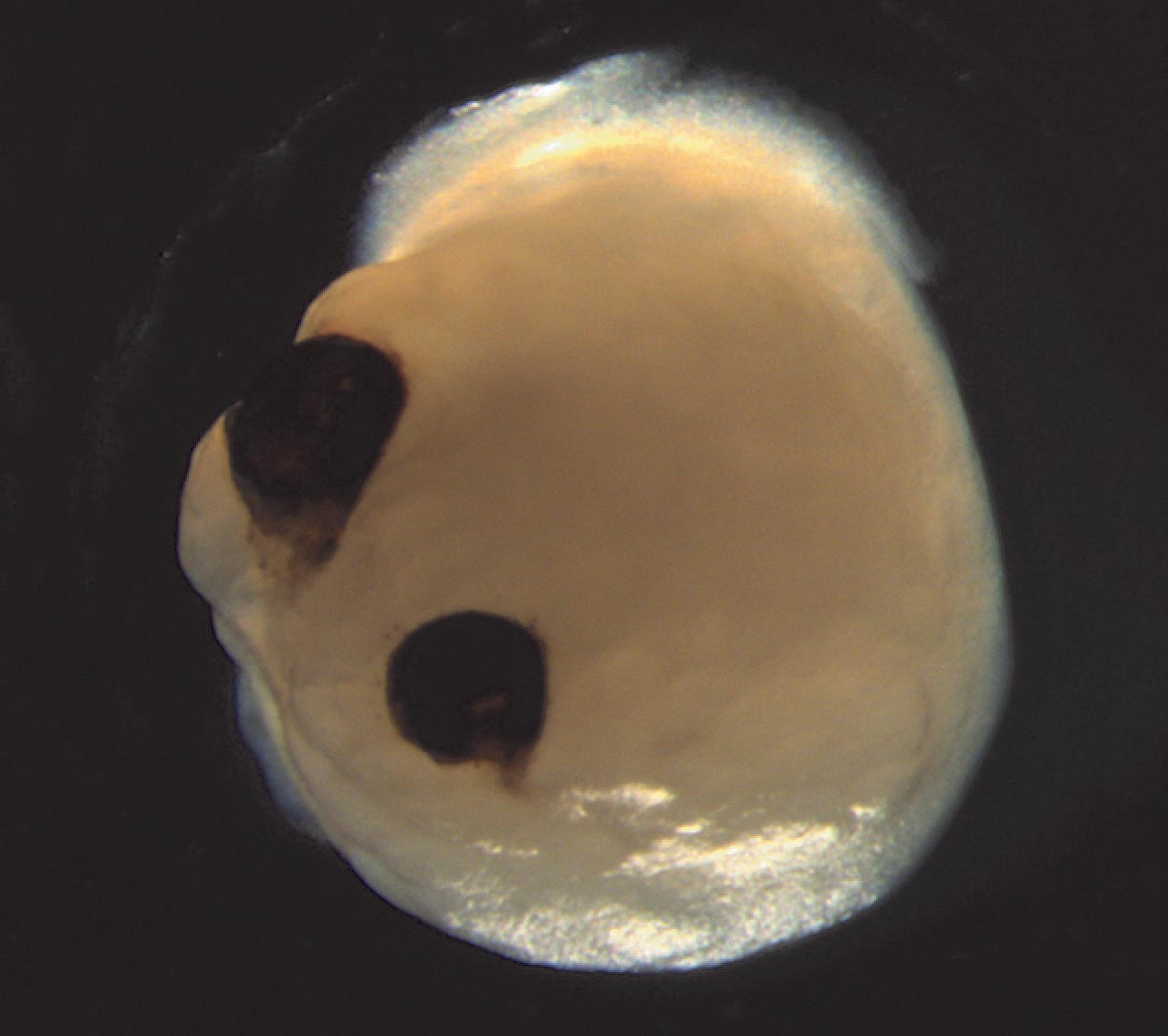
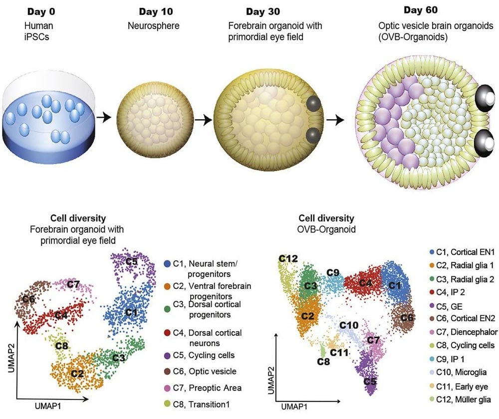
Human induced pluripotent stem cells (iPSCs) can be used to generate brain organoids containing an eye structure called the optic cup, according to a study published on August 17, 2021, in the journal Cell Stem Cell. The organoids spontaneously developed bilaterally symmetric optic cups from the front of the brain-like region, demonstrating the intrinsic self-patterning ability of iPSCs in a highly complex biological process.
“Our work highlights the remarkable ability of brain organoids to generate primitive sensory structures that are light sensitive and harbor cell types similar to those found in the body,” says senior study author Jay Gopalakrishnan of University Hospital Düsseldorf. “These organoids can help to study brain-eye interactions during embryo development, model congenital retinal disorders, and generate patient-specific retinal cell types for personalized drug testing and transplantation therapies.”
Many aspects of human brain development and diseases can be studied using 3D brain organoids derived from pluripotent stem cells, which can give rise to all cell types in the body. Researchers previously used human embryonic stem cells to generate the optic cup, which gives rise to the retina—the light-sensitive layer of tissue at the back of the eye. Another study demonstrated that optic-cup-like structures can be generated from iPSCs, which are derived from adult cells that have been genetically reprogrammed back into an embryonic-like pluripotent state.
In the past, the production of optic cups from pluripotent stem cells focused on generating the pure retina. Until now, optic cups and other 3D retinal structures had not been functionally integrated into brain organoids.
To achieve this feat, Gopalakrishnan and his team modified a protocol they previously developed for turning iPSCs into neural tissue. The human brain organoids formed optic cups, which appeared as early as 30 days and matured as visible structures within 50 days. This time frame parallels that of retinal development in the human embryo and could make certain types of developmental neurobiology experiments more efficient.
First Evidence of Retinal-Brain Connectivity In Vitro
Across 16 independent batches from four iPSC donors, the researchers generated 314 brain organoids, 72% of which formed optic cups, showing that the method is reproducible. These structures contained diverse retinal cell types, which formed electrically active neuronal networks that responded to light. The optic cup brain organoids also contained lens and corneal tissue and exhibited retinal connectivity to brain regions. “In the mammalian brain, nerve fibers of retinal ganglion cells reach out to connect with their brain targets, an aspect that has never before been shown in an in vitro system,” Gopalakrishnan says.
In future studies, they plan to develop strategies to keep the optic cups viable for long time periods, using them to investigate mechanisms that cause retinal disorders.
Reference: “Human brain organoids assemble functionally integrated bilateral optic vesicles” byElke Gabriel, Walid Albanna, Giovanni Pasquini, Anand Ramani, Natasa Josipovic, Aruljothi Mariappan, Friedrich Schinzel, Celeste M. Karch, Guobin Bao, Marco Gottardo, Ata Alp Suren, Jürgen Hescheler, Kerstin Nagel-Wolfrum, Veronica Persico, Silvio O. Rizzoli, Janine Altmüller, Maria Giovanna Riparbelli, Giuliano Callaini, Olivier Goureau, Argyris Papantonis, Volker Busskamp, Toni Schneider and Jay Gopalakrishnan, 17 August 2021, Cell Stem Cell.
DOI: 10.1016/j.stem.2021.07.010
Never miss a breakthrough: Join the SciTechDaily newsletter.
Follow us on Google and Google News.