
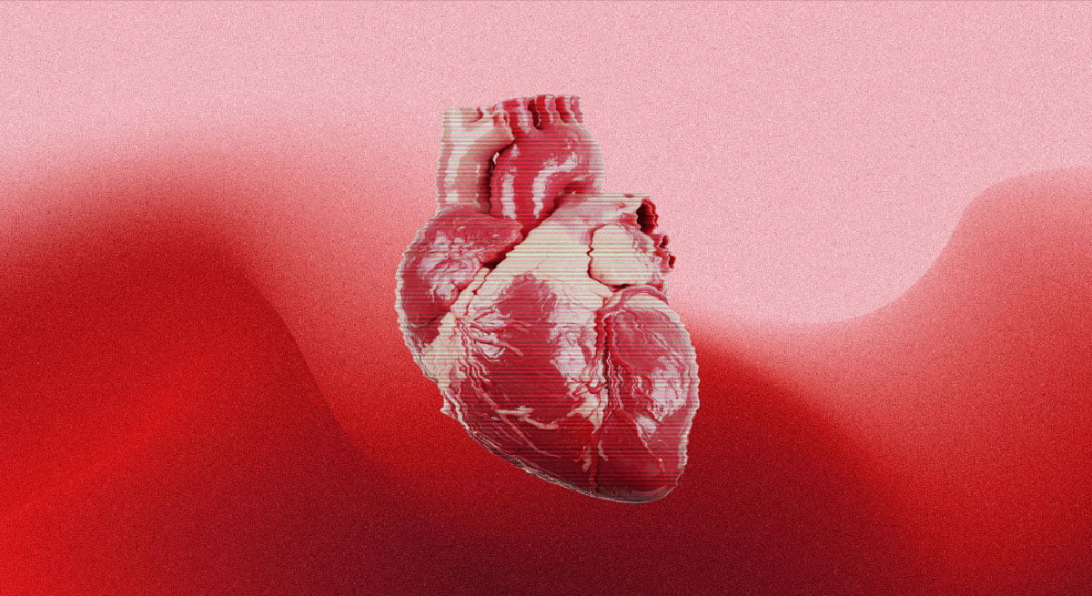
Researchers at the University of Copenhagen found that a signaling mechanism in the cells’ ‘antennae’ may help explain why some fetuses develop disease in both the heart and other organs.
About two of every 100 babies worldwide are born with congenital heart disease, but the biological processes responsible for these conditions are not fully understood.
Researchers at the University of Copenhagen have identified a previously unknown communication mechanism on the surface of cells that may provide part of the explanation.
“We have discovered a new communication system on the exterior of the cell that is crucial for the proper formation of the heart during embryonic development. This finding changes our understanding of how congenital heart defects arise. You could say that we have identified an important cog in a highly complex machine,” says Lars Allan Larsen, an expert in congenital heart disease and Professor at the Department of Cellular and Molecular Medicine.
The system operates within the primary cilium, a microscopic structure resembling an ‘antenna’ that extends from most cells in the body. Primary cilia interpret signaling molecules and help cells decide whether to divide, move, or die.
Three proteins guide heart formation
The researchers found that three proteins, TAK1, TAB2, and PKA-Cα, form a signaling center inside this cellular antenna and contribute significantly to the heart’s development.
“These proteins act as molecular instructions that tell stem cells when and how to develop into heart muscle cells. However, genetic alterations can disrupt this communication, causing ‘antenna defects,’ which may lead to congenital heart defects,” explains Søren Tvorup Christensen, Professor of cell biology at the Department of Biology.
Multiple experiments trace the mechanism
To determine how the mechanism works, the researchers combined patient genetic data with experiments involving zebrafish, human cells, and mouse stem cells.
They began by examining genetic information from several thousand people with congenital heart defects to identify rare mutations. The researchers looked for genetic changes that appeared more often among patients than healthy people, indicating a possible connection to the disease.
They then tested what those mutations did in a living organism. Through genetic engineering, the researchers reproduced the same changes in zebrafish and monitored how their hearts developed. The altered genes caused developmental abnormalities and impaired heart function in the fish.
The researchers also studied several laboratory-grown cell types to identify the molecular processes involved and determine what occurs when the relevant signaling pathways break down.
Together, the patient data and experimental results directed the researchers to the primary cilium, revealing a possible biological pathway behind congenital heart defects.
“We investigate the mechanism from many different angles and using many different methods, all of which support what we observe in patients. Therefore, we are reasonably confident that this mechanism also exists in humans,” says Lars Allan Larsen.
One disruption may affect several organs
The apparent effects extended beyond heart development.
The rare mutations were found in patients with syndromic congenital heart disease, in which an underlying genetic syndrome produces heart defects as well as abnormalities and related conditions in other organs.
Zebrafish experiments and detailed analyses of cilia in other tissues also indicated that the same signaling mechanism contributes to the development of multiple organs.
“When the ciliary mechanism fails, it typically affects the development of several other organs as well. This may explain why some patients with congenital heart disease also have defects and related conditions affecting the brain, kidneys, and skeleton. The mechanism provides a unifying explanation for diseases that we have previously struggled to understand,” says Søren Tvorup Christensen.
The discovery may therefore improve scientific understanding of several diseases caused by faulty primary cilia and could eventually contribute to new treatment approaches.
“Many rare genetic diseases are caused by changes in genes that affect ciliary function, yet the underlying mechanisms have remained poorly understood. This new knowledge may eventually make it easier to identify patients early and develop targeted treatments,” says Lars Allan Larsen.
Reference: “TAK1 operates at the primary cilium in non-canonical TGFB/BMP signaling to control heart development” by Canan Doganli, Oskar Kaaber Thomsen, Daniel A. Baird, Yeasmeen Ali, Menachem V. K. Sarusie, Enrique Audain, Line Jeanett Jessen, Pauline Munck Truelsen, Johanne Bay Mogensen, Maria Schrøder Holm, Kateřina Apolínová, Lorenzo Buttò, Maria Diamanti, Jindřiška Leischner Fialová, Emma M. Wade, Stephen P. Robertson, Lotte Bang Pedersen, Laurent Argiro, Fabienne Lescroart, Marc-Phillip Hitz, Søren Tvorup Christensen and Lars Allan Larsen, 4 August 2026, PLOS Biology.
DOI: 10.1371/journal.pbio.3003902
Never miss a breakthrough: Join the SciTechDaily newsletter.
Follow us on Google and Google News.