Researchers at King’s College London have identified significant differences in brain connectivity between term and pre-term infants, revealing that these early patterns are predictive of later developmental milestones.
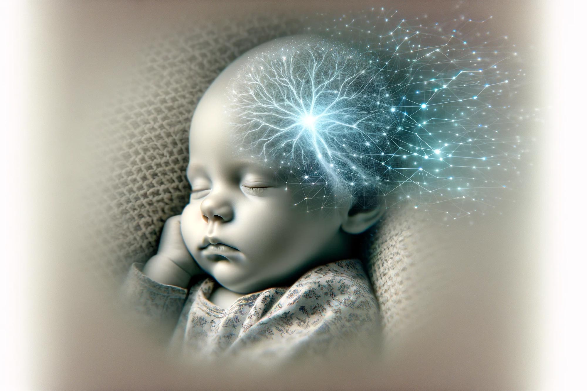
A new King’s College London scanning study of 390 babies has shown distinct patterns between term and pre-term babies in the moment-to-moment activity and connectivity of brain networks.
Supported by Wellcome and the National Institute of Health and Care Research (NIHR) Maudsley Biomedical Research Centre, this is the first study to analyze how the communication between brain areas changes moment-to-moment in the first few weeks of life.
Published today (February 8) in Nature Communications, the study also found that these dynamic patterns of brain connectivity in babies were linked to developmental measures of movement, language, cognition, and social behavior 18 months later.
Insights Into Early Brain Development
Joint senior author, Dr. Dafnis Batallé, Senior Lecturer in Neurodevelopmental Science at the Institute of Psychiatry, Psychology & Neuroscience (IoPPN), King’s College London said:
“Although we know how influential brain connectivity is on development, we know little about the patterns of dynamic functional connectivity in early life, and how they link to the way our brains mature. By analyzing brain scans from 390 babies, we have begun to identify different transient states of connectivity that could potentially provide insight into how the brain is developing at this age and what behaviors and functions these patterns are linked to as the baby grows older.”
There is increasing awareness that conditions such as ADHD, autism, and schizophrenia have their origins early in life, and that the development of these conditions may be linked to neonatal brain connectivity and its fluctuations over time.
Innovative Research Methods and Findings
Researchers used state-of-the-art techniques to evaluate functional Magnetic Resonance Imaging (fMRI) data on 324 full-term babies and 66 pre-term babies (born at less than 37 weeks gestation). They assessed how the connectivity changed moment-to-moment during the time the baby was in the scanner to provide a dynamic picture. Previous research with babies has always used a measure of connectivity averaged over time spent in the scanner.
Dr. Lucas França, first author and Assistant Professor in Computer and Information Sciences at Northumbria University. said:
“These findings are a result of carefully adapting methodologies derived from the domains of computer science and physics, specifically employed to unveil the intricacies inherent to the human neonatal brain. When these methodologies are allied to advanced techniques to obtain unprecedented data like the one from the Developing Human Connectome Project, we have a unique opportunity to deepen our understanding of the largely unknown realm of brain dynamics in early life.”
The study used methods that tap into how brain connectivity fluctuates: one method that considers connectivity patterns across the whole brain and one that considers patterns within different regions of the brain.
The study identified six different brain states: three of these were across the whole brain and three were constrained to regions of the brain (occipital, sensorimotor, and frontal regions). By comparing term and pre-term babies the researchers showed that different patterns of connectivity are linked to pre-term birth, for example, pre-term babies spent more time in frontal and occipital brain states than term babies. They also demonstrated that brain state dynamics at birth are linked to a range of developmental outcomes in early childhood.
Joint senior author, Professor Grainne McAlonan, Interim Director of NIHR Maudsley BRC and Professor of Translational Neuroscience at IoPPN, King’s College London said:
“This is a real step forward in the use of imaging techniques to investigate how brain activity is continually changing in early life and how this provides a platform to support subsequent developmental milestones in childhood. The difference between term and pre-term babies suggests that time spent in or outside the womb shapes brain development. We now need to try and find out if it is possible to use these insights to identify and help those who need some additional support.”
The data was sourced from The Developing Human Connectome Project (dHCP), which is led by King’s College London and funded by the European Research Council. It provides high-resolution magnetic resonance brain images from unborn and newborn babies to scientists worldwide to support many world-leading research projects into brain development and cerebral or mental health disorders.
Professor David Edwards, Principal Investigator of dHCP and Head of Department of Perinatal Imaging and Health, King’s College London said: “This study shows the power of the large set of data acquired by the Developing Human Connectome Project, an open science program funded by the European Research Council and led by King’s College London in collaboration with Imperial College London and the University of Oxford. The data are freely available to researchers who want to study human brain development.”
Reference: “Neonatal brain dynamic functional connectivity in term and preterm infants and its association with early childhood neurodevelopment” by Lucas G. S. França, Judit Ciarrusta, Oliver Gale-Grant, Sunniva Fenn-Moltu, Sean Fitzgibbon, Andrew Chew, Shona Falconer, Ralica Dimitrova, Lucilio Cordero-Grande, Anthony N. Price, Emer Hughes, Jonathan O’Muircheartaigh, Eugene Duff, Jetro J. Tuulari, Gustavo Deco, Serena J. Counsell, Joseph V. Hajnal, Chiara Nosarti, Tomoki Arichi, A. David Edwards, Grainne McAlonan and Dafnis Batalle, 8 February 2024, Nature Communications.
DOI: 10.1038/s41467-023-44050-z
Never miss a breakthrough: Join the SciTechDaily newsletter.
Follow us on Google and Google News.
2 Comments
This study strongly suggests the fetal brain continues changing until birth. It implies there is some significant change, a reason to draw a line “between pre-term and term babies”. But the line it finds could be the result of medical interventions, and not a line that indicates a significant change at birth. Obstetricians will sometimes artificially induce labor and ther circumstances, such as injury, cann cause early labor. And of course there is caesarean birth. All these potential disruptions themselves force the categorical distinction between pre-term and term. Once that distinction is made, of course it would necessarily create a categorical distinction in ANY measure of brain function. A distinction would be seen no matter how “sophisticated”, “Innovative”, “state-of-the-art”, “dynamic”, “adapting”, advanced”, “unprecedented”, “a real step forward”, or “high-resolution” (adjectives from the article) the measurement is. The only way for a negative result in any measurement would be if the fetal brain doesn’t change at all past viability. Apparently the researchers DID NOT attempt to correct for the division artificially created by Obstetricians, etc. Perhaps by incorporating gestational time as a variable, and placing the babies on a continuum, instead of accepting the division as a given. The “differences” measured in babies brains may be a continuous, first-order (linear) change, which would mean there is no significant change at birth. We already knew the post-natal brain changes continuously. I think the only people who might believe the fetal brain didn’t also change continuously are the forced-birther people. The continuous change before birth and after may also connect continuously. In other words, birth may not cause a discontinuity in any measure of brain development. The researchers failure to de-categorize, so to speak, the pre-categorized data makes their study unable to speak to this question, and they cannot claim to find new “significant differences”, other than the fact that the brain is continuously changing until and after birth. No matter how sophisticated one’s instruments, a particular measurement schema can mislead when the much sought-after measurability is prioritized over repeatability.
The information is valuable.