Scientists are expanding plankton cells to reveal the hidden architecture that powers life in the oceans.
Plankton quietly sustain life on Earth. These microscopic organisms generate a large portion of the oxygen we breathe and sit at the base of the ocean food web. They are also extraordinarily diverse, with tens of thousands of known species and countless others still unknown. Within this vast group, protists, tiny single-celled organisms, are especially important for understanding evolution. Yet for decades, scientists were limited to studying them through genetic information alone, because dependable ways to see their internal structures did not exist.
A Pandemic Call Sparks a Breakthrough
During the COVID-19 pandemic, EMBL Group Leader Gautam Dey received a Zoom call from his collaborator Omaya Dudin, who was then leading a research group at EPFL. Dudin had recently modified a new method that made it possible to visualize the internal structure of Ichthyosporea — a marine protist closely related to animals and fungi — solving a long-standing problem caused by its impenetrable cell walls.
The approach, known as expansion microscopy, was originally developed by scientists at MIT, USA. It was later refined into ultrastructure expansion microscopy (U-ExM) by Paul Guichard and Virginie Hamel at the University of Geneva to study sub-cellular ultrastructure. This improved technique made the cell walls permeable, allowing researchers to clearly observe and analyze the protist’s internal components.
From Collaboration to a Global Vision
Encouraged by these results, Dudin, Dey, Guichard, and Hamel launched a close collaboration. Three years later, their work has produced near-encyclopedic insight into hundreds of protist species and is moving toward an ambitious goal: creating a planetary atlas of plankton.
A major boost came from the EMBL-led Traversing European Coastlines (TREC) expedition, which allowed the team to explore the internal organization of marine microbes in far greater depth. Their findings, recently published in Cell, provide detailed views of the cellular architecture of more than 200 plankton species, especially eukaryotes – organisms whose cells contain a nucleus enclosed by a membrane. This research marks the launch of PlanExM, a TREC plug-in project designed to uncover planktonic ultrastructural diversity using expansion microscopy.
A Treasure Trove of Marine Microorganisms
One of the first major sampling sites on the TREC expedition was Roscoff, France. There, the Station Biologique maintains one of Europe’s most extensive collections of marine microorganisms. The researchers asked the facility’s manager, Ian Probert, how many samples might be available for a pilot study with expansion microscopy. They expected around 20 species, but instead were granted access to more than 200.
“We spent three days and nights just fixing those samples. This was a treasure trove we could not let go of,” said co-first author Felix Mikus, who completed his PhD in the Dey Group and is now a postdoc in Dudin’s lab at the University of Geneva.
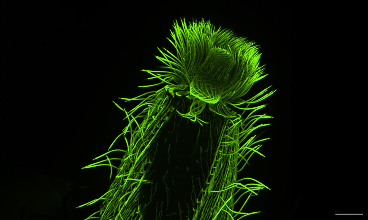
How Expansion Microscopy Makes the Invisible Visible
Expansion microscopy is still a young technique, turning 10 years old this year, and it works by physically enlarging biological samples. A sample – which may include single-celled organisms, tissues, or individual cells – is embedded in a transparent gel. When the gel absorbs water, it expands. Many internal structures remain intact and stretch proportionally, effectively enlarging the sample by four or even 16 times without relying on powerful lenses.
“When combined with regular light microscopy methods, expansion microscopy allows scientists to bypass the standard wavelength barriers which limit how small a structure can be resolved using light microscopy,” said Guichard and Hamel.
Mapping the Cell’s Internal Framework
Using samples from Roscoff and another culture collection from Bilbao, Spain, the researchers carried out one of the most extensive studies to date on cytoskeletal diversity. The cytoskeleton is the filament network that gives eukaryotic cells their structure and organization. The team focused on microtubules – hollow tubes that help cells maintain shape, divide, and move – as well as centrins, a group of proteins involved in organizing microtubules within the cell.
“We were able to map features of microtubule and centrin organization across many different eukaryotic groups,” said Hiral Shah, an EIPOD Postdoctoral Fellow in EMBL’s Dey and Schwab groups and co-first author of the study. “The scale of the study, with many species characterized in each group, opens up the possibility to make evolutionary predictions. For instance, dinoflagellates, one of the most diverse groups found in oceans across the planet, are well-represented in our study. We were able to map the presence of tubulin and centrin structures associated with the cell cortex or the flagella in these species.”
Linking Cell Structure to Evolution
“U-ExM is transforming how we explore protist ultrastructure,” said Armando Rubio Ramos, co-first author of the study and a Postdoctoral Fellow in the Hamel and Guichard research group at the University of Geneva. “By combining this technique with high-throughput imaging and comparative analyses, we can begin to decode how cellular architecture has diversified across evolution. It’s a bridge between molecular data and the physical organization of life at the microscopic scale.”
The researchers say this work sheds light on the basic principles that govern how eukaryotic cells are built, while also offering clues about how cytoskeletal structures evolved over time. It also highlights the strength of expansion microscopy as a tool for analyzing complex samples, including those taken directly from marine environments.
Toward a Planetary Atlas of Plankton
“Our adventures with expansion microscopy are only beginning,” said Dey. “This is perhaps the first high-resolution microscopy technique that has the potential to match the scale and ambition of large biodiversity genomics projects, enabling us in the near future to associate new multiomics data with cellular physiology at scale across the tree of life.”
With Oxford University’s Thomas Richards joining the effort, Dey and Dudin also secured a CHF 2 million Moore Foundation Grant to continue the research.
“The next step is to selectively look deeper into certain species within this broad collection to answer specific questions, such as understanding how mitosis and multicellularity evolved and the phenotypic diversity that underlie major evolutionary transitions,” Dudin said.
Reference: “Charting the landscape of cytoskeletal diversity in microbial eukaryotes” by Felix Mikus, Armando Rubio Ramos, Hiral Shah, Jonas Hellgoth, Marine Olivetta, Susanne Borgers, Clémence Saint-Donat, Margarida Araújo, Chandni Bhickta, Paulina Cherek, Jone Bilbao, Estibalitz Txurruka, Yana Eglit, Nikolaus Leisch, Yannick Schwab, Filip Husnik, Sergio Seoane, Ian Probert, Paul Guichard, Virginie Hamel, Gautam Dey and Omaya Dudin, 31 October 2025, Cell.
DOI: 10.1016/j.cell.2025.09.027
Never miss a breakthrough: Join the SciTechDaily newsletter.
Follow us on Google and Google News.