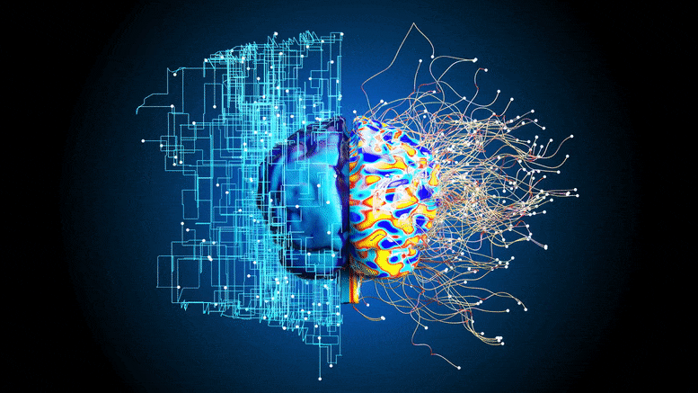
Neuroscientists at the Medical University of South Carolina use a novel brain imaging technique to visualize subtle brain changes in pre-symptomatic Alzheimer’s patients.
Neuroscientists from the Medical University of South Carolina (MUSC) report in the journal Brain Connectivity that they have detected subtle differences in the way the brain functions in older adults with preclinical Alzheimer’s disease (AD).
Adults with preclinical AD have the earliest signs of disease, such as the buildup of amyloid-beta proteins in their brains. However, they have no noticeable symptoms of cognitive decline.
“Using these individualized maps of brain function, we found a potential brain-based reason for very subtle cognitive changes in this early phase of the disease.” Dr. Andreana Benitez
The research team, led by Andreana Benitez, Ph.D., and Stephanie Fountain-Zaragoza, Ph.D., used a novel brain imaging analysis technique to construct individualized maps of brain function. They then looked to see if there were links between subtle changes in brain function and declining cognitive performance, assessed using behavior-based tests. This approach could improve the ability to study the preclinical phase of AD.
“Prior studies have not found an association between brain function and behavior in preclinical AD,” said Benitez. “Using these individualized maps of brain function, we found a potential brain-based reason for very subtle cognitive changes in this early phase of the disease.”
Detecting Subtle Changes in Brain Function With Improved Brain Mapping
Research into the preclinical phase of AD could help us to understand how the disease begins and progresses. However, early changes in brain function are very subtle, making them challenging to study. With pilot project funding from the South Carolina Clinical & Translational Research Institute, the MUSC researchers used a new form of brain mapping to detect these subtle effects.
They looked at brain activity using a functional connectome – a type of brain map that measures how different brain regions communicate with one another. Think of the brain like a big city, said Fountain-Zaragoza, where brain regions are clustered into neighborhoods connected by highways. The functional connectome is like watching the activity across that city – how much there is going on within each neighborhood and how well traffic flows between them.
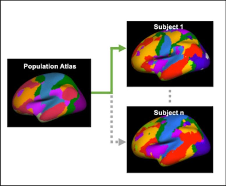
The researchers relied on a newer and highly sensitive form of image analysis to appreciate how these neighborhoods function in individuals. The technique – the individualized functional connectome – was developed by their collaborator Hesheng Liu, Ph.D.
Traditional functional connectomes use an average of many people’s brains as a map for functional brain regions. In contrast, Liu’s method can show the unique patterns of brain function for each individual.
“We all have the same functional parts of our brain, but they’re positioned slightly differently, sort of like a fingerprint,” said Fountain-Zaragoza. “This method creates an individualized brain fingerprint that more accurately reflects where the different functional regions are in each individual’s brain.”
Linking Subtle Changes in Brain Function to Behavior
The researchers used this novel brain fingerprinting technique to look for subtle changes in brain function in 149 participants aged 45 to 85 without signs of cognitive decline. All participants underwent PET scans of their brains and were divided into two groups – those with and without PET scan evidence of early amyloid-beta protein buildup. The participants also underwent MRI scans, which were used to generate the brain fingerprints.
“This method creates an individualized brain fingerprint that more accurately reflects where the different functional regions are in each individual’s brain.” Dr. Stephanie Fountain-Zaragoza
The researchers then tested how well the participants in each group performed on behavior-based tests of information processing. They found that certain changes in the brain fingerprint were associated with worse information processing in participants with amyloid-beta buildup, or preclinical AD.
In participants with preclinical AD, information processing was worse in those with greater than usual between-network connectivity, or too much activity on the brain’s highways. In contrast, information processing was better in those with higher within-network connectivity, or more brain activity within important neighborhoods of the brain.
“A healthy brain typically has a balance of connectivity within and between its networks,” said Fountain-Zaragoza. “We found that in preclinical AD—when amyloid build-up is present in the brain—this balance can be disrupted, potentially leading to information no longer being processed as efficiently.”
What the Study Teaches Us
The study shows that individualized functional connectomes can detect subtle variations in brain function that could be missed with other conventional brain imaging analysis techniques.
It also suggests that the early stages of amyloid-beta buildup could affect the function of brain networks even before symptoms of cognitive decline become noticeable.
Finally, it reveals that changes in connectivity within and between specific brain networks may indicate early problems with information processing. This imbalance in connectivity could therefore be a good target for therapies to improve the outcomes for patients with AD.
Next steps
With renewed grant funding from the National Institute on Aging, Benitez and Fountain-Zaragoza plan to continue their work on preclinical AD. They hope to focus more on the extent to which brain changes affect disease progression and also exploring new treatments, such as brain stimulation, which may help to slow it.
“There’s a lot of great work aimed at helping us understand the earliest signs and symptoms of Alzheimer’s disease,” said Fountain-Zaragoza. “This area of work is important for understanding the full spectrum of the disease and identifying who might be at risk of developing it.”
Reference: “Functional Network Alterations Associated with Cognition in Pre-Clinical Alzheimer’s Disease” by Stephanie Fountain-Zaragoza, Hesheng Liu and Andreana Benitez, 17 March 2023, Brain Connectivity.
DOI: 10.1089/brain.2022.0032
Funding: NIH/National Center for Advancing Translational Sciences (NCATS), NIH/National Institute on Aging
Never miss a breakthrough: Join the SciTechDaily newsletter.
Follow us on Google and Google News.
1 Comment
Conti’s audience engagement events bring a unique and enjoyable dimension to films.