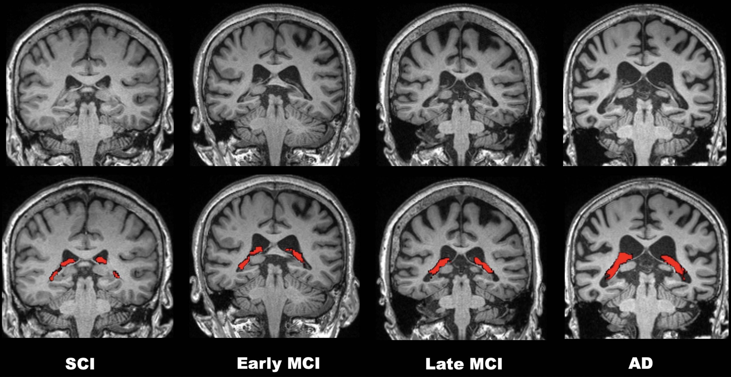
Increased volume of an important structure in the brain called the choroid plexus is linked to greater cognitive impairment and Alzheimer’s disease, according to a new study published today (May 17, 2022) in the journal Radiology.
The choroid plexus is a network of blood vessels, connective tissue, and cells found in spaces of the brain called ventricles. The plexus plays a critical role in brain health. It is a gateway for immune cells from the blood to the brain, and as the primary site for the production of cerebrospinal fluid, it is crucial to removing waste products and toxic proteins from brain cells. In the case of Alzheimer’s disease, this role is particularly important. Recent research suggests that disease progression is related to the accumulation of abnormal proteins called amyloid and tau and subsequent degeneration of the nerves.
“Researchers believe impaired clearance rather than overproduction of abnormal amyloid and tau is responsible for Alzheimer’s disease,” said study senior author Won-Jin Moon, M.D., Ph.D., professor of radiology and chairperson of the Department of Radiology at the Konkuk University School of Medicine in Seoul, Korea. “Thus, we assume that the abnormal status of choroid plexus is linked to the failure of clearance leading to waste and toxic protein accumulation in the brain and failure of immune surveillance leading to neuroinflammation.”
Little is known about the choroid plexus’ imaging profile in cognitive impairment.
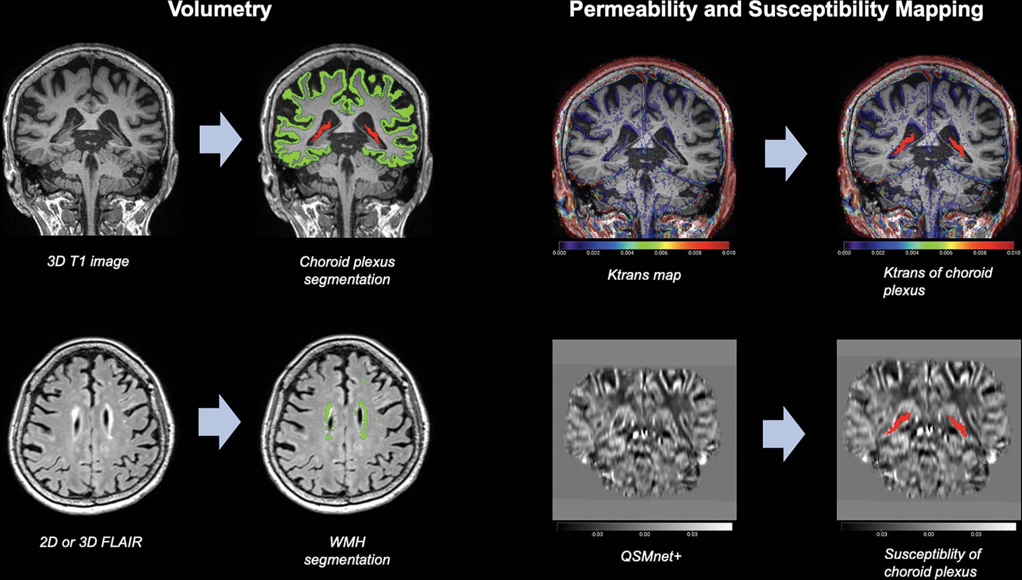
To learn more, Dr. Moon and colleagues performed brain MRI on 532 participants at various stages of cognitive impairment. Of the 532 participants, 132 underwent permeability imaging using dynamic contrast-enhanced MRI.
“Our study found that the enlarged choroid plexus volume is independently associated with increased cognitive impairment,” Dr. Moon said. “We found no relationship between choroid plexus volume and amyloid pathology but a clear relationship between the choroid plexus volume and cognitive impairment severity.”
The study results point to new possibilities for MRI’s role in the diagnosis of Alzheimer’s disease.
“I think our findings on the choroid plexus can suggest it as a new potential MR imaging surrogate for an impaired clearance system and neuroinflammation,” Dr. Moon said.
Other potential clinical applications include helping researchers develop new target drugs or treatments for clearance failure and neuroinflammation. Eventually, choroid plexus measurements could help speed treatment to those who need it most.
“If we combine choroid plexus volume and hippocampal volume in a screening stage, it may help us better discriminate the more vulnerable patients from the less vulnerable ones,” Dr. Moon said.
The researchers plan to follow up with a longitudinal study. They will explore changes in choroid plexus volume over time as the disease progresses.
Reference: “Choroid Plexus Volume and Permeability at Brain MRI within the Alzheimer Disease Clinical Spectrum” by Jong Duck Choi, Yeonsil Moon, Hee-Jin Kim, Younghee Yim, Subin Lee, Won-Jin Moon, 17 May 2022, Radiology.
DOI: 10.1148/radiol.212400
Never miss a breakthrough: Join the SciTechDaily newsletter.
Follow us on Google and Google News.