
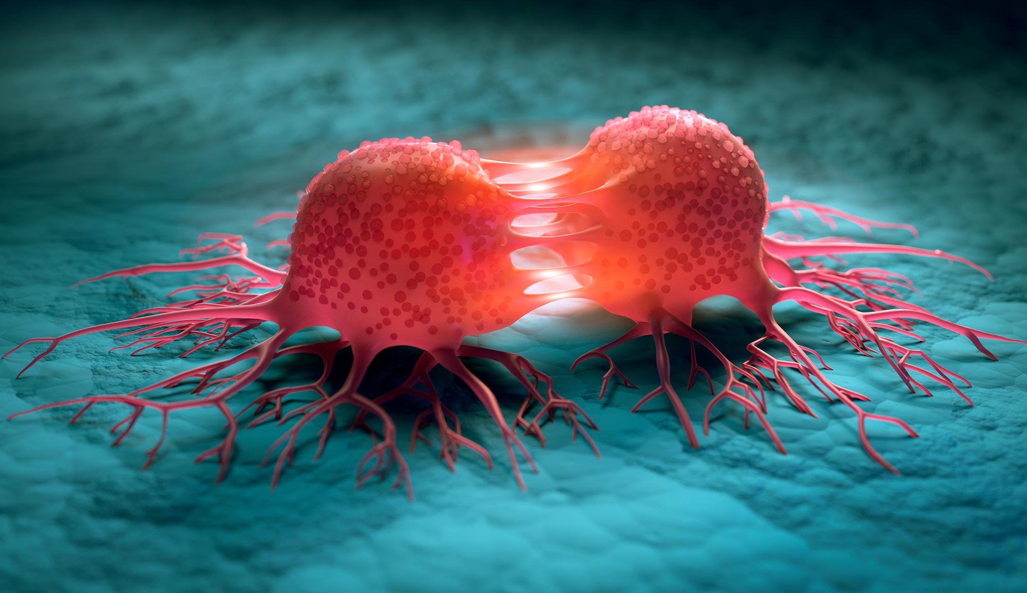
Cancer’s spread to distant organs can now be studied using lab-grown bone, lung, and blood vessel tissues.
Cancer becomes far more dangerous when it stops being confined to one place.
Most cancer deaths are not caused by the original tumor, but by cells that break away, travel through the bloodstream, and establish new tumors in distant organs. This process, known as metastasis, is responsible for at least two-thirds of cancer deaths. Yet despite decades of research, drugs designed specifically to prevent or halt metastatic progression have had limited success.
One major obstacle is the shortage of predictive human models that allow scientists to watch the process unfold and identify what makes particular organs vulnerable.
Animal research has revealed much about how cancer spreads, but rodent biology cannot fully reproduce what happens in patients. Those differences may help explain why treatments that appear promising in laboratory animals often disappoint in clinical trials.
A Human Model of Cancer Spread
Researchers at Columbia Engineering have now developed a multi-organ chip that recreates one of metastasis’s most elusive stages. The system follows circulating breast cancer cells as they leave vascular flow and enter engineered human bone and lung tissue. The study, led by Columbia Engineering professor Gordana Vunjak-Novakovic, was published in Science Translational Medicine.
The first model of its kind, the chip contains separate compartments with millimeter-scale bone and lung tissues connected by a flowing vascular channel. This design does more than place human tissues beside cancer cells. It allows researchers to observe the continuous exchange of signals between cells in the bloodstream and the organs they may eventually colonize.
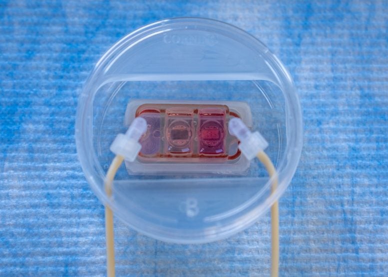
“The pressing need for developing human tissue models of metastasis has been a key motivation for our study,” said Vunjak-Novakovic, who is a university professor at Columbia University and the Mikati Foundation Professor of Biomedical Engineering and Professor of Medical Sciences (in Medicine). “Our objective was to probe the ability of cancer cells to adhere to and traverse across endothelium [inner lining of blood vessels] and to determine their capacity to survive in the tissues they are colonizing through cell reprogramming and niche remodeling.”
How Cancer Invades Distant Organs
Reaching another organ is only the beginning of metastasis. Cancer cells must attach to the blood vessel lining, cross that barrier, evade local defenses, and adapt to unfamiliar tissue. Only then can they multiply and form a secondary tumor. This phase, known as organ colonization, is exceptionally difficult to observe directly in patients or reproduce accurately in animals.
Metastasis is also selective rather than random. Different cancer cells tend to favor particular organs, a pattern known as “organ tropism.” Breast cancer commonly spreads to sites including bone and lung, making those tissues especially useful for testing whether the chip could reproduce behavior seen in the human body.
The platform also accepts patient cells and engineered human tissues, giving scientists a way to investigate individual differences in metastatic behavior. Researchers can alter one component at a time, examine organ-specific interactions, and search for molecular pathways that could become new treatment targets.
“Cancer is very smart, unfortunately. We learned how the cells cross barriers to get from blood circulation into the tissues,” said Vunjak-Novakovic. “We were also able to reproduce something that happens in patients, where cancer cells condition the target tissues, even before they colonize them, to make them more receptive.”
Building Bone, Lung, and Blood Vessels
The team produced bone, lung, and vascular endothelium from induced pluripotent stem cells (iPSCs), which can be guided to develop into many specialized cell types. Tissue-specific scaffolds and bioreactors helped each tissue mature, while separate compartments provided the conditions needed to preserve its function over time. Vascular circulation then linked the compartments.
A selectively permeable endothelial barrier separated the flowing channel from the organ tissues, just as blood vessel walls do inside the body. The researchers introduced breast cancer cells into the circulation and watched as they encountered the barrier, crossed into nearby tissue, and established distinct patterns of colonization.
The results closely reflected the cells’ known preferences. Breast cancer cells that typically spread to bone colonized the engineered bone more strongly and produced more extensive degeneration. Cells with an affinity for lung tissue caused greater damage there while colonizing the bone only modestly. Differences in tissue invasion and secreted molecules showed that the device captured key features of organ-specific metastasis.
Cancer Prepares the Ground
The chip also reproduced a more insidious feature of cancer spread. Before metastatic cells fully occupy a distant organ, they can release signals that alter its environment and make it easier to invade. Scientists call this preparation of future tumor sites pre-metastatic niche formation.
Analysis of both tissue compartments revealed evidence that the breast cancer cells were conditioning distant organs before colonization. Recreating this early transformation could help researchers investigate opportunities to interrupt metastasis before a secondary tumor becomes established.
“The key advantages of this advanced model of metastasis are that it is human and can be patient-specific,” Vunjak-Novakovic said. “It faithfully mimics some of the key aspects of human metastasis that are otherwise largely inaccessible for direct study.”
A New Direction for Preclinical Research
Vunjak-Novakovic leads Columbia’s Laboratory for Stem Cells and Tissue Engineering, which was among the early developers of microphysiological systems, often called “organs on chips.” These small human tissue platforms are designed to model disease and drug responses under more realistic conditions than conventional cell cultures.
The project also brought together researchers from Columbia’s Herbert Irving Comprehensive Cancer Center, including Andrea Califano, the Clyde and Helen Wu Professor of Chemical and Systems Biology; Peter Sims, associate professor of systems biology; and Hanina Hibshoosh, professor of pathology & cell biology.
Rather than replacing animal research outright, engineered human tissues could fill important gaps and help scientists determine which discoveries are most likely to translate to patients. Ilaria Baldassarri, a Columbia Engineering PhD student and one of the study’s lead authors, said the platform demonstrates how that emerging approach could work in practice.
“As the FDA and NIH place growing emphasis on new approach methodologies, this study is a concrete example of what that shift can look like in practice, applied to one of cancer’s most challenging hallmarks: metastasis,” Baldassarri said.
Reference: “Organ-specific colonization and niche remodeling in a human tissue model of metastasis” by Alan G. Chramiec, Ilaria Baldassarri, Ece Öztürk, Daniel Naveed Tavakol, Miranda C. Wang, Max Summers, Keith Yeager, Richard Z. Zhuang, Anushka Srivastava, Somnath Tagore, Diogo Teles, Hanina Hibshoosh, Andrea Califano, Peter A. Sims and Gordana Vunjak-Novakovic, 19 August 2026, Science Translational Medicine.
DOI: 10.1126/scitranslmed.adv6871
This study was funded by the NIH/National Cancer Institute and the Chan Zuckerberg Initiative.
Never miss a breakthrough: Join the SciTechDaily newsletter.
Follow us on Google and Google News.