
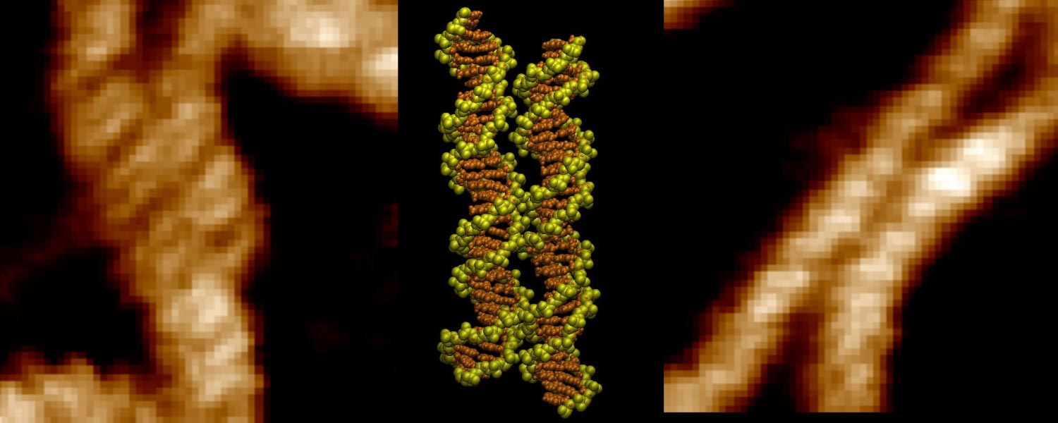
Scientists have revealed how tiny metal ions help DNA molecules overcome electrical repulsion and “zip” together with striking precision.
DNA molecules carry the same negative charge, so they would normally be expected to push apart. Inside living cells, however, DNA must sometimes come together for processes such as genetic recombination, gene silencing and cancer development.
Researchers have now observed how that pairing can happen with striking precision. Using high-powered atomic force microscopy, the team watched short DNA fragments align groove for groove. Computer simulations then showed that positively charged metal ions can settle into those grooves and act as tiny molecular bridges that help hold the two DNA molecules together.
Tiny Ions Help DNA Molecules Pair
Professor Agnes Noy, from the School of Physics, Engineering and Technology at the University of York, co-led the research. She said: “This discovery could help researchers identify regions of the genome specially involved in DNA pairing. These regions may become particularly important when mutations disrupt normal cellular processes and contribute to cancer.”
The results support a theory proposed about 20 years ago known as the “DNA zipper” model. Professor Alexey Kornyshev from Imperial College London and his collaborators originally suggested that salt ions surrounding DNA could create alternating patterns of charge. Those patterns would allow neighboring DNA molecules to align in a way resembling interlocking spiral staircases.
Testing the “DNA Zipper” Model
To investigate the idea, the researchers scanned DNA samples with atomic force microscopy, producing detailed maps of their surfaces. At the same time, sophisticated computer simulations followed the behavior of individual atoms and ions.
The simulations revealed that double-charged metal ions can behave almost like a pair of charged arms. By interacting with both DNA molecules at once, the ions help bridge the space between them and stabilize their alignment.
Dr. Thomas Catley, co-lead author from the School of Chemical Materials and Biological Engineering at the University of Sheffield, said: “It was incredible to be able to directly visualize the long-hypothesized mechanism for the first time. The advanced imaging techniques at our disposal are allowing us to uncover these key DNA interactions which have implications in many key cellular processes.”
Dr. Victor Velasco-Berrelleza from the University of Sheffield, who performed the simulations, added: “Microscopy shows us what happens, but the simulations allow us to uncover the molecular mechanism behind it.”
DNA Pairing Hotspots
The researchers also found that DNA molecules do not all pair with the same strength. Some DNA sequences form much stronger contacts than others, creating hotspots where two helices are especially likely to line up.
These sequence-dependent interactions could help scientists identify regions of the genome that are particularly important for DNA pairing. That may be valuable for understanding how mutations interfere with normal cellular behavior and contribute to cancer.
The findings could also have applications beyond medicine. Because some DNA sequences can be designed to interact more strongly than others, the same principles may eventually help engineers build custom DNA structures for biotechnology.
Reference: “Imaging and mechanism of DNA–DNA recognition mediated by divalent ions” by Thomas E Catley, Victor Velasco-Berrelleza, Daniel E Rollins, Alice L B Pyne and Agnes Noy, 25 August 2026, Nucleic Acids Research.
DOI: 10.1093/nar/gkag817
Never miss a breakthrough: Join the SciTechDaily newsletter.
Follow us on Google and Google News.