
A tiny, flexible electronic device that wraps around the spinal cord could represent a new approach to the treatment of spinal injuries, which can cause profound disability and paralysis.
A team of engineers, neuroscientists, and surgeons from the University of Cambridge developed the devices and used them to record the nerve signals going back and forth between the brain and the spinal cord. Unlike current approaches, the Cambridge devices can record 360-degree information, giving a complete picture of spinal cord activity.
Enhancing Spinal Injury Treatment
Tests in live animal and human cadaver models showed the devices could also stimulate limb movement and bypass complete spinal cord injuries where communication between the brain and spinal cord had been completely interrupted.
Most current approaches to treating spinal injuries involve both piercing the spinal cord with electrodes and placing implants in the brain, which are both high-risk surgeries. The Cambridge-developed devices could lead to treatments for spinal injuries without the need for brain surgery, which would be far safer for patients.
Benefits and Long-term Potential
While such treatments are still at least several years away, the researchers say the devices could be useful in the near-term for monitoring spinal cord activity during surgery. Better understanding of the spinal cord, which is difficult to study, could lead to improved treatments for a range of conditions, including chronic pain, inflammation, and hypertension. The results are reported today (May 8) in the journal Science Advances.
“The spinal cord is like a highway, carrying information in the form of nerve impulses to and from the brain,” said Professor George Malliaras from the Department of Engineering, who co-led the research. “Damage to the spinal cord causes that traffic to be interrupted, resulting in profound disability, including irreversible loss of sensory and motor functions.”
The ability to monitor signals going to and from the spinal cord could dramatically aid in the development of treatments for spinal injuries, and could also be useful in the nearer term for better monitoring of the spinal cord during surgery.
Advanced Monitoring Technologies
“Most technologies for monitoring or stimulating the spinal cord only interact with motor neurons along the back, or dorsal, part of the spinal cord,” said Dr. Damiano Barone from the Department of Clinical Neurosciences, who co-led the research. “These approaches can only reach between 20 and 30 percent of the spine, so you’re getting an incomplete picture.”
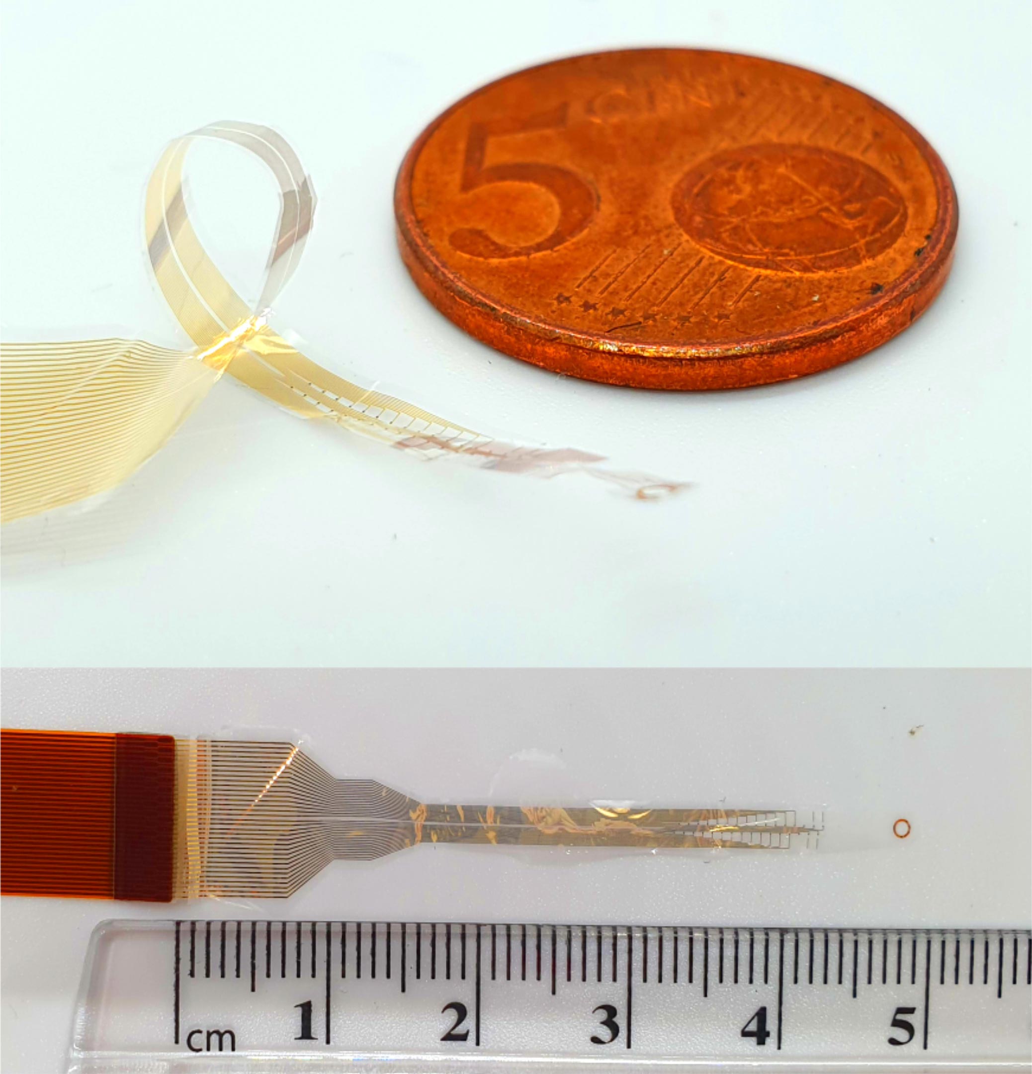
By taking their inspiration from microelectronics, the researchers developed a way to gain information from the whole spine, by wrapping very thin, high-resolution implants around the spinal cord’s circumference. This is the first time that safe 360-degree recording of the spinal cord has been possible – earlier approaches for 360-degree monitoring use electrodes that pierce the spine, which can cause spinal injury.
The Cambridge-developed biocompatible devices – just a few millionths of a meter thick – are made using advanced photolithography and thin film deposition techniques, and require minimal power to function.
The devices intercept the signals traveling on the axons, or nerve fibers, of the spinal cord, allowing the signals to be recorded. The thinness of the devices means they can record the signals without causing any damage to the nerves, since they do not penetrate the spinal cord itself.
“It was a difficult process, because we haven’t made spinal implants in this way before, and it wasn’t clear that we could safely and successfully place them around the spine,” said Malliaras. “But because of recent advances in both engineering and neurosurgery, the planets have aligned and we’ve made major progress in this important area.”
Implementation and Future Prospects
The devices were implanted using an adaptation to routine surgical procedure so they could be slid under the spinal cord without damaging it. In tests using rat models, the researchers successfully used the devices to stimulate limb movement. The devices showed very low latency – that is, their reaction time was close to human reflexive movement. Further tests in human cadaver models showed that the devices can be successfully placed in humans.
The researchers say their approach could change how spinal injuries are treated in the future. Current attempts to treat spinal injuries involve both brain and spinal implants, but the Cambridge researchers say the brain implants may not be necessary.
“If someone has a spinal injury, their brain is fine, but it’s the connection that’s been interrupted,” said Barone. “As a surgeon, you want to go where the problem is, so adding brain surgery on top of spinal surgery just increases the risk to the patient. We can collect all the information we need from the spinal cord in a far less invasive way, so this would be a much safer approach for treating spinal injuries.”
While a treatment for spinal injuries is still years away, in the nearer term, the devices could be useful for researchers and surgeons to learn more about this vital, but understudied, part of human anatomy in a non-invasive way. The Cambridge researchers are currently planning to use the devices to monitor nerve activity in the spinal cord during surgery.
“It’s been almost impossible to study the whole of the spinal cord directly in a human, because it’s so delicate and complex,” said Barone. “Monitoring during surgery will help us to understand the spinal cord better without damaging it, which in turn will help us develop better therapies for conditions like chronic pain, hypertension, or inflammation. This approach shows enormous potential for helping patients.”
Reference: “Flexible circumferential bioelectronics to enable 360-degree recording and stimulation of the spinal cord” by Ben J. Woodington, Jiang Lei, Alejandro Carnicer-Lombarte, Amparo Güemes-González, Tobias E. Naegele, Sam Hilton, Salim El-Hadwe, Rikin A. Trivedi, George G. Malliaras and Damiano G. Barone, 8 May 2024, Science Advances.
DOI: 10.1126/sciadv.adl1230
The research was supported in part by the Royal College of Surgeons, the Academy of Medical Sciences, Health Education England, the National Institute for Health Research, and the Engineering and Physical Sciences Research Council (EPSRC), part of UK Research and Innovation (UKRI).
Never miss a breakthrough: Join the SciTechDaily newsletter.
Follow us on Google and Google News.