Cells have their own hidden “wind system” that rapidly moves proteins and could change cancer research.
Researchers at Oregon Health & Science University have identified a previously unknown system inside cells that works like internal “trade winds,” rapidly carrying essential proteins to the cell’s leading edge. This discovery is changing how scientists understand cell movement, cancer spread, and wound healing.
The findings, published in I, challenge long-standing ideas about how proteins get to the right place at the right time inside cells.
For decades, scientists believed that proteins floating within cells moved mainly by diffusion, drifting randomly until they reached their destination. The new research shows that this process is not left to chance. Cells actively generate directed fluid flows that push key proteins forward to areas where movement and repair take place.
Unexpected Classroom Experiment Leads to Breakthrough
The discovery began with an unplanned observation during a neurobiology course at the Marine Biological Laboratory in Massachusetts. The study’s co-corresponding authors, Catherine (Cathy) Galbraith, Ph.D., and James (Jim) Galbraith, Ph.D., were running a routine experiment with students when something unusual appeared.
“It actually started out as an unexpected finding,” Cathy said. “We were just conducting an experiment with students in class.”
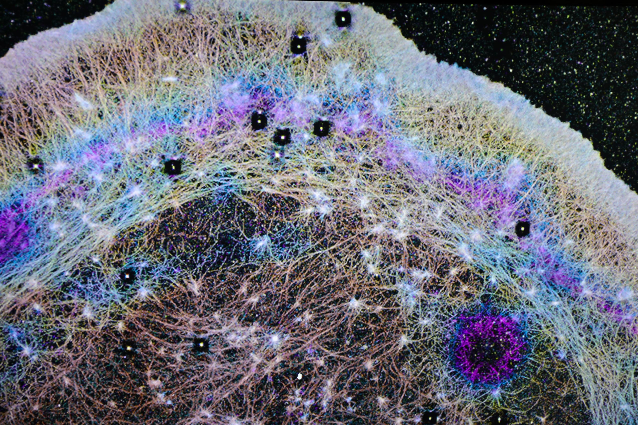
In the experiment, a laser was used to make proteins temporarily invisible in a strip at the back of a living cell, a common method for tracking movement inside cells. During the process, the researchers noticed a second dark line forming at the front edge of the cell, the area that extends as the cell moves.
“We kind of did it for fun and then realized this gave us a way of measuring something that wasn’t able to be measured before,” she said.
Further analysis revealed that this dark band represented a wave of soluble actin, a protein that plays a central role in cell movement, being pushed rapidly to the front. Previously, scientists thought actin mainly arrived there by random diffusion. The new results showed a completely different mechanism at work.
“We realized the cartoon models in textbooks were missing a huge piece,” Jim said. “There had to be some kind of flow in the cell pushing things forward. Cells really do ‘go with the flow.’”
Directed Fluid Flows Drive Cell Movement
Cathy and Jim joined OHSU in 2013 after working at the National Institutes of Health, where they collaborated with Nobel Laureate Eric Betzig, Ph.D., at the Howard Hughes Medical Institute’s Janelia Research Campus to develop advanced live cell imaging tools.
Using specialized imaging assays, the team found that cells actively create directional fluid flows, similar to atmospheric rivers. These currents move actin and other proteins toward the front of the cell far more quickly than diffusion alone.
“We found that the cell can actually squeeze at the back and target where it sends that material,” Jim said. “If you squeeze half a sponge, the water only goes on that half. That’s basically what the cell is doing.”
These flows are nonspecific, meaning they carry many different proteins at once. This creates a fast and efficient transport system that supports cell protrusion, adhesion and rapid shape changes. These processes are essential for cell movement, immune responses, and tissue repair.
The researchers also found that the flows occur within a specialized region at the cell’s front. This area is separated from the rest of the cell by an actin-myosin condensate barrier, which acts like a physical boundary that directs proteins to advancing parts of the cell edge.
New Imaging Technique Reveals Cellular Currents
To observe these internal movements, the team developed a modified version of a standard fluorescence technique. Instead of removing fluorescence, they activated fluorescent molecules at a single point and tracked how they spread.
One of their key experiments was named FLOP, or Fluorescence Leaving the Original Point.
“It wasn’t a flop at all,” Cathy said. “It was the opposite. It is anything but a flop, because it worked.” The team’s discovery may help explain why certain cancer cells move so aggressively.
Insights Into Cancer Cell Migration
The findings offer a possible explanation for why some cancer cells are especially invasive.
“We know these highly invasive cells have this really cool mechanism to push proteins really fast, really rapidly where they need them at the front of the cell,” Jim said. “All cells have basically the same components inside, much like a Porsche and a Volkswagen have many of the same parts, but when those parts are assembled into the final machine, they behave and function very differently.”
By identifying how this system operates differently in cancer cells, researchers hope to find new ways to slow or stop their movement.
“If you can understand the differences, you can target future therapies based on how cancer cells and normal cells work differently,” he said.
Collaboration and Advanced Microscopy Made It Possible
This project combined expertise from engineering, physics, microscopy, and cell biology. Key collaborators at Janelia Research Campus in Virginia contributed advanced tools, including fluorescence correlation spectroscopy and 3D super resolution imaging.
“The instrumentation we needed doesn’t exist in most places,” Cathy said. “Janelia had a one-of-a-kind setup that let us test and confirm what we were seeing.”
The work relied heavily on cutting-edge imaging methods developed at Janelia, including iPALM, an interferometric technique that can resolve structures at the nanometer scale.
“iPALM allowed us to physically see the compartments,” Jim said. “There’s no other light-based technique that could do that.”
Discovery of a New “Pseudo Organelle”
The researchers describe this system as a “pseudo organelle,” a functional compartment that is not enclosed by a membrane but still plays a major role in organizing cell activity.
“Just as small shifts in the jet stream can change the weather, small changes in these cellular winds could change how diseases begin or progress,” Cathy said.
The team believes this discovery could open new possibilities in cancer research, drug delivery, tissue repair and synthetic biology.
“All you had to do was look,” Cathy said. “The flows were there all along. Now we know how cells use them.”
Reference: “Compartmentalized cytoplasmic tradewinds direct soluble proteins” by Catherine G. Galbraith, Brian P. English, Ulrike Boehm and James A. Galbraith, 30 March 2026, Nature Communications.
DOI: 10.1038/s41467-026-70688-6
In addition to the Galbraiths, coauthors on this study are Brian English, Ph.D., of Janelia Research Campus, and Ulrike Boehm, Ph.D., formerly with Janelia and now with Carl Zeiss AG of Germany.
This study was supported by the National Institute of General Medical Sciences, of the National Institutes of Health, under Award number R01GM117188, U.S. National Science Foundation, under Award numbers 2345411 and 171636, the W. M. Keck Foundation, the Howard Hughes Medical Institute Janelia Visiting Scientist Program, and the Howard Hughes Medical Institute. The iPALM work was partly supported by an award from the Advanced Imaging Center at Janelia. The SIM imaging was partly supported by a Core Research Facilities grant from OHSU School of Medicine.
Never miss a breakthrough: Join the SciTechDaily newsletter.
Follow us on Google and Google News.