Six feet of human DNA crammed into a tiny nucleus relies on an elegant system of nucleosomes, fibers, and highly organized phase-separated condensates.
Scientists have now captured the most detailed images yet of how chromatin fibers and nucleosomes arrange themselves inside these droplet-like structures, revealing how molecular architecture determines condensate behavior.
How Cells Fit Six Feet of DNA Into Tiny Nuclei
Inside every human cell, biology manages an extraordinary challenge: packing roughly six feet of DNA into a nucleus that is only about one-tenth the width of a human hair, all while keeping the genetic material fully functional.
To achieve this level of compression, DNA coils around proteins to form nucleosomes. These nucleosomes connect like beads on a string, creating long strands that fold into chromatin fibers. The fibers then compact even further to fit inside the nucleus.
For years, scientists did not know exactly how this final stage of compaction occurred. That changed in 2019, when HHMI Investigator Michael Rosen and his colleagues at UT Southwestern Medical Center showed that lab-made nucleosomes can gather into membrane-less droplets called condensates. They discovered that this occurs through phase separation – a process similar to oil droplets forming in water – which may mirror how chromatin becomes densely packed within living cells.
Exploring the Hidden Behavior of Chromatin Condensates
Chromatin condensates contain hundreds of thousands of rapidly moving molecules. When these molecules come together, they display emergent properties that individual components do not possess on their own. These group behaviors determine how droplets form, hold together, and retain their physical traits.
To understand these properties in detail and learn how chromatin compacts inside cells, researchers needed a close view of the inner arrangement of nucleosomes and chromatin fibers within the droplets.
Rosen’s team, collaborating with HHMI Investigator Elizabeth Villa at the University of California, San Diego; Rosana Collepardo-Guevara at the University of Cambridge; and Zhiheng Yu at HHMI’s Janelia Research Campus, has now accomplished this.
Using advanced imaging technologies at Janelia, the scientists produced the most detailed visualizations to date of the molecular layout inside synthetic chromatin condensates. The same methods were then applied to examine chromatin inside actual cells.
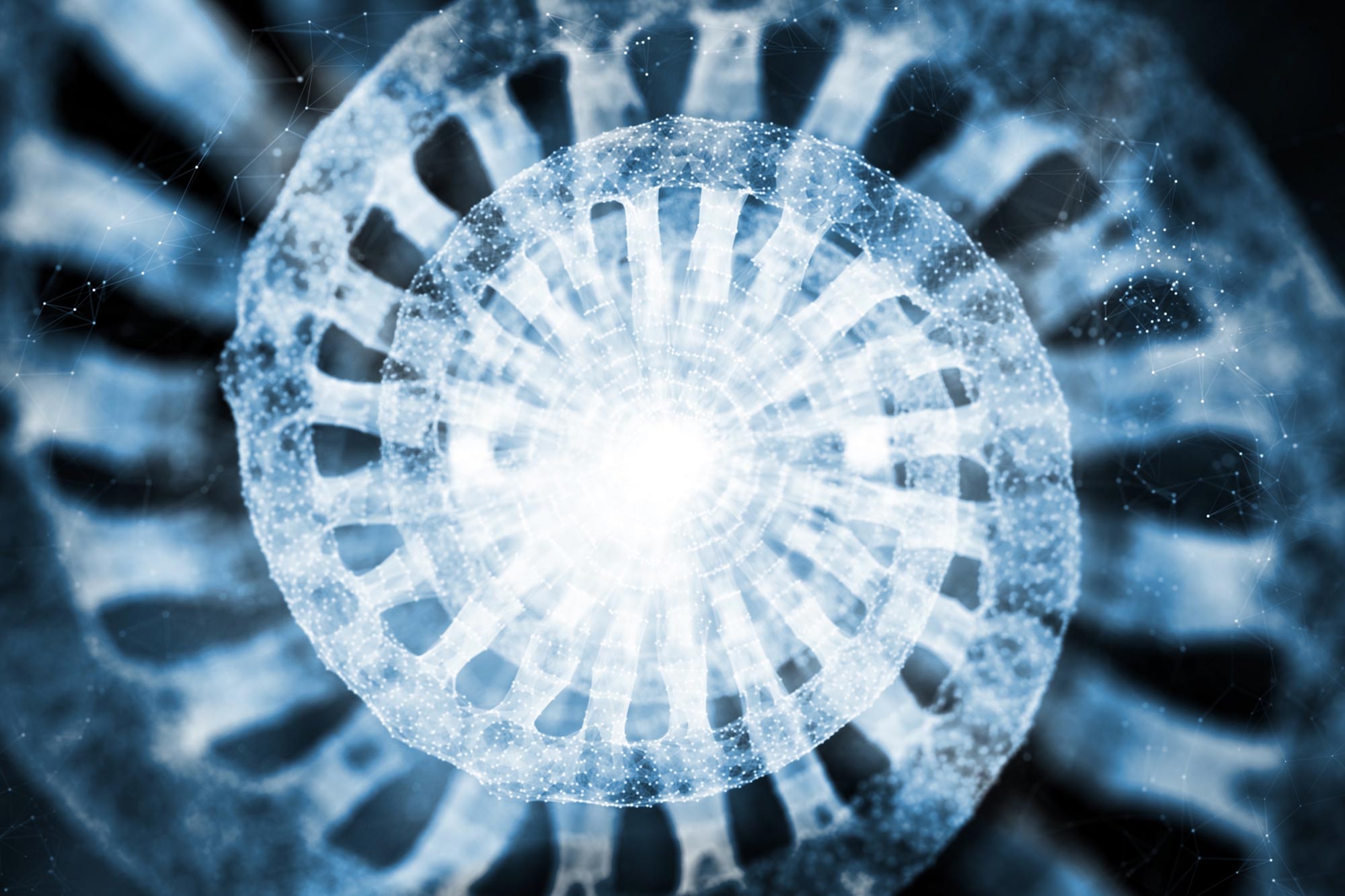
A team of researchers led by HHMI Investigator Michael Rosen has captured the most detailed images yet of the molecules in synthetic chromatin condensates — droplet-like structures of compacted DNA. These movies show images of the chromatin condensate obtained by cryo-electron tomography, revealing the individual nucleosomes making up the droplet, followed by a high-resolution reconstruction of the nucleosomes in the chromatin condensate. Credit: Zhou et al.
What High-Resolution Imaging Reveals About Condensate Structure
By combining these images with computer simulations and light microscopy, the research team was able to analyze how individual molecules within the droplets are arranged and how they interact. This helped them begin to understand the rules that govern how condensates form and function.
One key finding was that the length of the linker DNA between nucleosomes influences the overall organization of the structures. The spacing determines how chromatin fibers interact and shapes the larger network inside the condensates.
These structural features clarified why some types of chromatin undergo phase separation more readily than others and why condensates made from different chromatin forms exhibit distinct material properties. The team also discovered that synthetic condensates produced in the laboratory closely resemble the tightly packed chromatin found inside living cells.
“The work has allowed us to tie the structures of individual molecules to macroscopic properties of their condensates, really for the first time,” Rosen says. “I’m certain that we’re only at the tip of the iceberg – that we and others will come up with even better ways of developing those structure-function relationships at the meso (intermediate) scale.”
A Framework for Understanding Biomolecular Condensation
This research extends beyond chromatin. The new insights provide a model for studying a wide range of biomolecular condensates, which play key roles across the cell in processes such as gene regulation and stress response.
Learning how these droplet-like structures assemble and operate may also clarify what happens when condensation breaks down. Such failures are thought to contribute to several health conditions, including neurodegenerative diseases and cancer.
“By doing this research, we will better understand how abnormal condensation could lead to different diseases and, potentially, that could help us develop a new generation of therapeutics,” says Huabin Zhou, a postdoctoral scientist in the Rosen Lab and the lead author of the new research.
Reference: “Multiscale structure of chromatin condensates explains phase separation and material properties” by Huabin Zhou, Jan Huertas, M. Julia Maristany, Kieran Russell, June Ho Hwang, Run-Wen Yao, Nirnay Samanta, Joshua Hutchings, Ramya Billur, Momoko Shiozaki, Xiaowei Zhao, Lynda K. Doolittle, Bryan A. Gibson, Andrea Soranno, Margot Riggi, Jorge R. Espinosa, Zhiheng Yu, Elizabeth Villa, Rosana Collepardo-Guevara and Michael K. Rosen, 4 December 2025, Science.
DOI: 10.1126/science.adv6588
Never miss a breakthrough: Join the SciTechDaily newsletter.
Follow us on Google and Google News.
2 Comments
nice topic
Very good Allah technology