A new study presents a minimally invasive, ultra-flexible electronic neural implant, delivered via blood vessels, which records single-neuron activity in deep-brain regions, offering promising advancements for brain-machine interfaces and personalized neural therapies.
Introduction to Ultra-Small Neural Implants
A revolutionary ultra-small, ultra-flexible electronic neural implant has been developed that can record single-neuron activity deep within rat brains, as detailed in a new study.
“This technology could enable long-term, minimally invasive bioelectronic interfaces with deep-brain regions, writes Brian Timko in a related Perspective.
Brain-Machine Interfaces and Their Limitations
Brain-machine interfaces (BMIs) facilitate direct electrical communication between the brain and external electronic systems. These devices allow brain activity to directly control objects like prostheses, or modulate nerve or muscle function, thus aiding individuals with paralysis or neurological disorders to regain function.
However, the majority of conventional BMIs are only able to measure neural activity at the brain’s surface. In order to record single-neuron activity from deeper brain regions, invasive intracranial surgery is often necessary to implant probes. Such procedures can lead to complications including infection, inflammation, and damage to brain tissues.
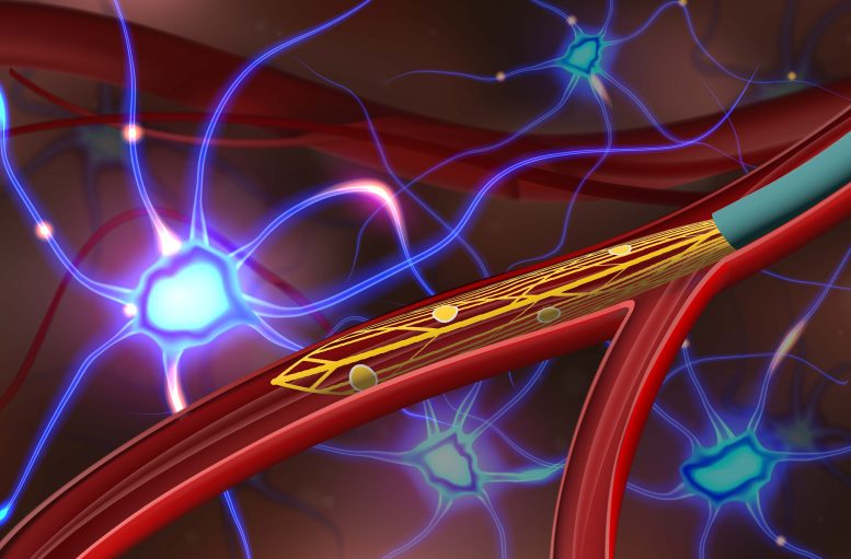
Micro-Endovascular Probes: A Less Invasive Approach
An alternative to the invasive surgical implantation of bioprobes into deep-brain regions is the utilization of the brain’s vascular network. In this study, Anqi Zhang and colleagues introduce ultra-flexible micro-endovascular (MEV) probes that can be accurately delivered to deep-brain regions via blood vessels. The team designed an ultra-small, flexible, mesh-like electronic recording device that can be placed onto a flexible microcatheter and implanted into blood vessels of less than 100-micron scale in the inner brain.
Testing and Validation of MEV Probes
Upon delivery, this device expands like a stent to record neuronal signals across the vascular wall without causing harm to the brain or its vasculature. To evaluate the potential of the MEV probe in vivo, the researchers implanted this injectable probe into the vasculature of rat brains. They demonstrated its ability to measure local field potentials and single-neuron activity in the cortex and olfactory bulb. Further, the researchers found that the implanted devices showed long-term stability, did not significantly alter cerebral blood flow or rat behavior, and triggered only a minimal immune response.
Implications and Future Developments
Brian Timko points out that future versions of such devices could provide customized therapies for patients by recording and decoding their neural activity and subsequently providing the appropriate modulatory stimuli.
Reference: “Ultraflexible endovascular probes for brain recording through micrometer-scale vasculature” by Anqi Zhang, Emiri T. Mandeville, Lijun Xu, Creed M. Stary, Eng H. Lo and Charles M. Lieber, 20 July 2023, Science.
DOI: 10.1126/science.adh3916
Never miss a breakthrough: Join the SciTechDaily newsletter.
Follow us on Google and Google News.