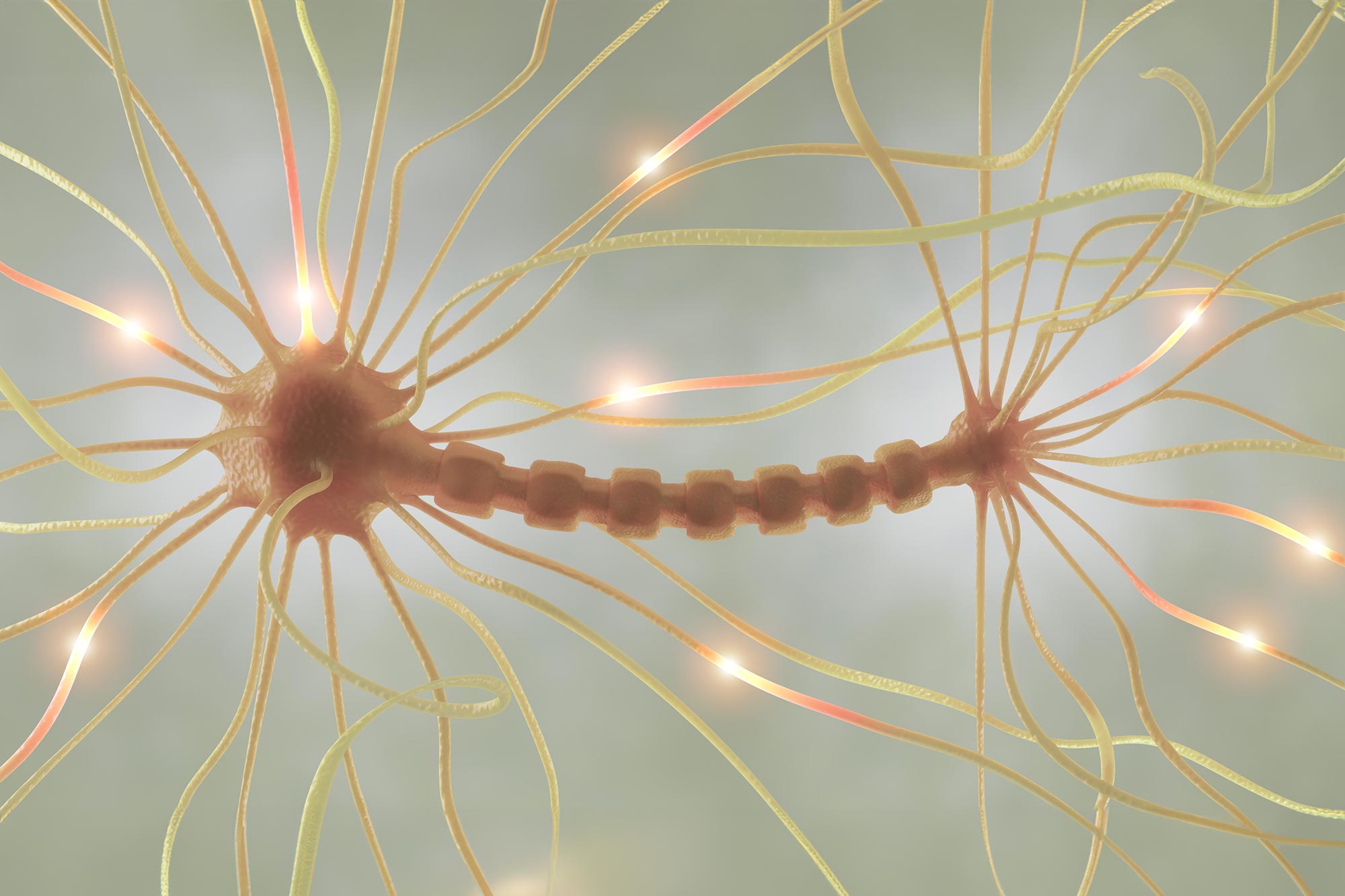
New research challenges traditional views on axon structure, presenting a “pearls-on-a-string” model where action potential speed is influenced by nano-pearl size changes due to local factors like cholesterol.
This finding, observed in cryo-preserved mouse nervous tissues, could impact understanding of neurodegenerative diseases linked to cholesterol regulation.
Rethinking Axon Structure
In a provocative study, scientists are challenging a long-held belief in neuroscience about the structure of axons—thin, elongated fibers that transmit electrical signals between nerve cells. The research, led by Shigeki Watanabe of Johns Hopkins School of Medicine and partially conducted at the Marine Biological Laboratory (MBL) Neurobiology course, introduces a new model for understanding how information flows in the brain. The findings are published today (December 5) in Nature Neuroscience.
For over 70 years, axons have been depicted as ultrathin, cylindrical cables that vary slightly in diameter but maintain a mostly uniform shape. Electrical signals, or action potentials, were thought to travel through axons at a steady speed, much like cars moving smoothly through a tunnel. This concept stems from the pioneering work of Alan Hodgkin and Andrew Huxley in the 1940s and 1950s, which included studies on the squid giant axon conducted at MBL.
Axons: Pearls-on-a-String Morphology Unveiled
However, Watanabe and team demonstrate that axons actually have a “pearls-on-a-string” morphology at the nanoscale level – lengths of cable interspersed with bulges they call “nano-pearls” (or nonsynaptic boutons). The speed of the action potential isn’t constant, they assert, but modulated by changes in the size of the nano-pearls, which in turn are caused by mechanical changes in the axon’s membrane and cytoskeleton as the action potential travels through.
“We can think of this like cars traveling on a highway,” Watanabe says. “If you have a four-lane highway through a tunnel, the cars travel normally. But if the highway has four lanes, then narrows to one lane, then goes back to four lanes, one lane – that’s how axons actually look. And you’d think the flow of traffic wouldn’t be too great.”
“But what’s interesting is the size of the pearls-on-a-string can change, at certain locations,” Shigeki continues. “We’ve shown you can modulate the nano-pearls’ size by changing factors in the local area, such as cholesterol in the plasma membrane. That, in turn, modulates the speed of the action potential. So, axons are highly flexible in that sense.”

Flash-Freezing Before Imaging Led to Discovery
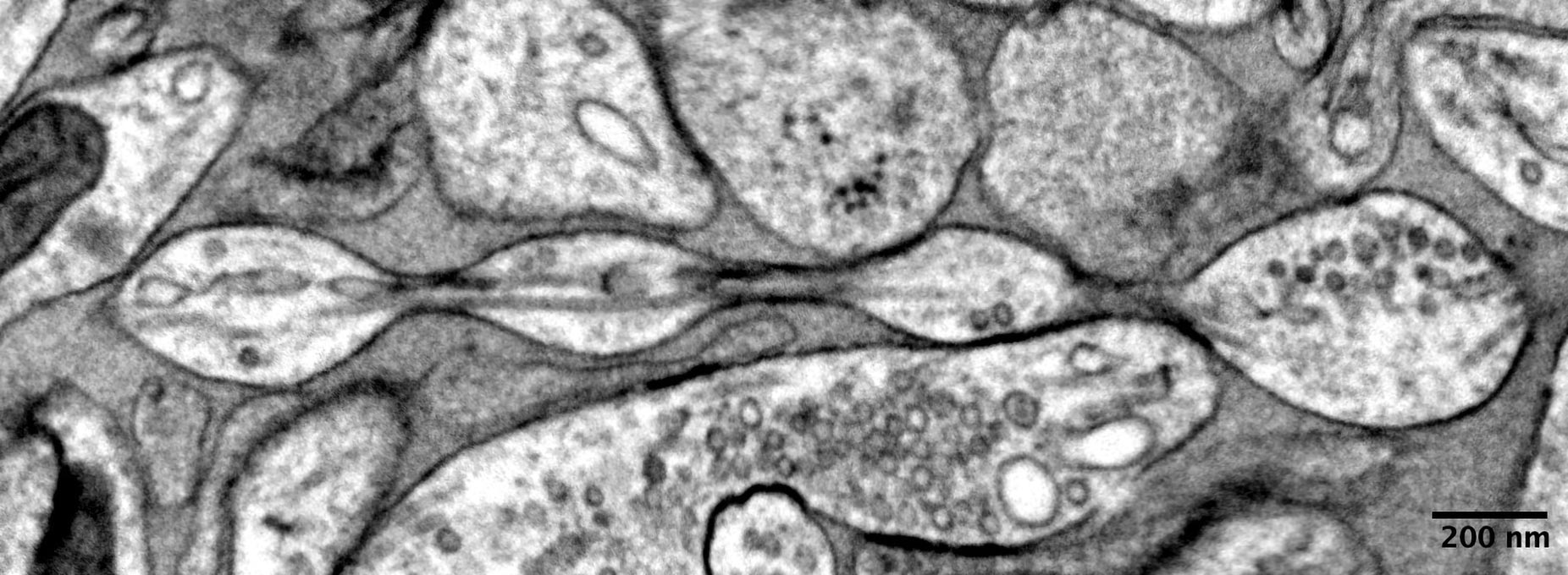
This axon ultrastructure that Watanabe and team describe is far below the diffraction limit of light microscopy, with the axon tract being about 60 nm in diameter with repeated nano-pearls about 200 nm in diameter. (The observations were made in unmyelinated axons in a mouse nervous system.)
“The reason people have missed this axon morphology before, and that we were able to observe this, is because we are looking at cryo-preserved tissues under an electron microscope,” Watanabe said. “Usually, people use chemicals to process samples for electron microscopy and then dehydrate these tissues, which is like making a grape into a raisin. But when you cryo-preserve, it’s like you’re making a frozen grape. You can preserve the actual shape.”
Implications for Neurodegenerative Diseases
This discovery has implications for understanding neurodegenerative disease, Watanabe said. Alzheimer’s disease, for example, is associated with misregulation of cholesterol in the brain. Watanabe’s study shows the size of nano-pearls is modified by cholesterol moving onto or out of the neuronal plasma membrane, which in turn regulates the conduction velocity of action potentials. If this mechanism is impaired, it may eventually lead to axonal death.
“It will be interesting in the future to look at the mutations that lead to neurodegeneration, what axon morphology looks like in those neurons, and whether axon plasticity is still present,” he said.
Reference: “Membrane mechanics dictate axonal pearls-on-a-string morphology and function” by Jacqueline M. Griswold, Mayte Bonilla-Quintana, Renee Pepper, Christopher T. Lee, Sumana Raychaudhuri, Siyi Ma, Quan Gan, Sarah Syed, Cuncheng Zhu, Miriam Bell, Mitsuo Suga, Yuuki Yamaguchi, Ronan Chéreau, U. Valentin Nägerl, Graham Knott, Padmini Rangamani and Shigeki Watanabe, 2 December 2024, Nature Neuroscience.
DOI: 10.1038/s41593-024-01813-1
Since 2015, Watanabe has been on the faculty of the MBL Neurobiology course, where part of the research was conducted. First author Jacqueline Griswold and co-authors Siyi Ma and Renee Pepper are MBL Neurobiology course alumni.
Part of this work was supported by an MBL Whitman Fellowship to Watanabe.
Never miss a breakthrough: Join the SciTechDaily newsletter.
Follow us on Google and Google News.
1 Comment
Seems spike currents running through the sinuous sheath could critically be creating electrostrictive oscillations.