A groundbreaking study has mapped a human cerebral cortex fragment in unprecedented detail, finding crucial cellular details and providing a tool for future research.
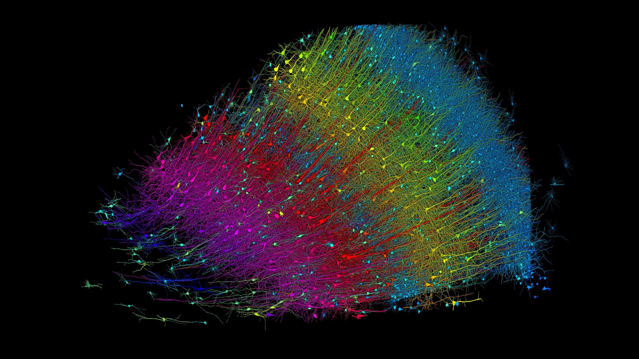
Using more than 1.4 petabytes of electron microscopy (EM) imaging data, researchers have generated a nanoscale-resolution reconstruction of a millimeter-scale fragment of human cerebral cortex, providing an unprecedented view into the structural organization of brain tissue at the supracellular, cellular, and subcellular levels. The human brain is a vastly complex organ and, to date, little is known about its cellular microstructure, including the synaptic and neural circuits it supports. Disruption of these circuits is known to play a role in myriad brain disorders.
Challenges in Brain Research
Studying human brain samples at such a high level of detail presents numerous challenges. These include technological limitations and the difficulties in obtaining and preserving tissue samples from healthy individuals. In their groundbreaking work, Alexander Shapson-Coe and colleagues performed a high-resolution EM reconstruction of the ultrastructure of a cubic millimeter of human temporal cortex. The reconstruction includes roughly 57,000 cells, about 230 millimeters of blood vessels, and nearly 150 million synapses, making up 1400 terabytes of data.
Insights from the Data
The scientists generated a three-dimensional reconstruction of nearly every cell and process within the cubic millimeter sample. They also developed a freely available tool for visualizing and analyzing the vast dataset. Using the data, the authors uncovered previously underappreciated aspects of the human temporal cortex, such as the disproportionate number of glia over neurons and the presence of rare yet potent axonal inputs with up to ~50 synapses each.
“Further studies using this resource may bring valuable insights into the mysteries of the human brain,” write the researchers.
For more on this research, see Harvard and Google Create Intricately Detailed 1,400 Terabyte 3D Brain Map.
Reference: “A petavoxel fragment of human cerebral cortex reconstructed at nanoscale resolution” by Alexander Shapson-Coe, Michał Januszewski, Daniel R. Berger, Art Pope, Yuelong Wu, Tim Blakely, Richard L. Schalek, Peter H. Li, Shuohong Wang, Jeremy Maitin-Shepard, Neha Karlupia, Sven Dorkenwald, Evelina Sjostedt, Laramie Leavitt, Dongil Lee, Jakob Troidl, Forrest Collman, Luke Bailey, Angerica Fitzmaurice, Rohin Kar, Benjamin Field, Hank Wu, Julian Wagner-Carena, David Aley, Joanna Lau, Zudi Lin, Donglai Wei, Hanspeter Pfister, Adi Peleg, Viren Jain and Jeff W. Lichtman, 10 May 2024, Science.
DOI: 10.1126/science.adk4858
Never miss a breakthrough: Join the SciTechDaily newsletter.
Follow us on Google and Google News.