
Researchers have visualized plant-to-plant communication via airborne compounds, identifying the specific signals and cellular responses that activate plant defenses against threats.
Airborne Communication Among Plants
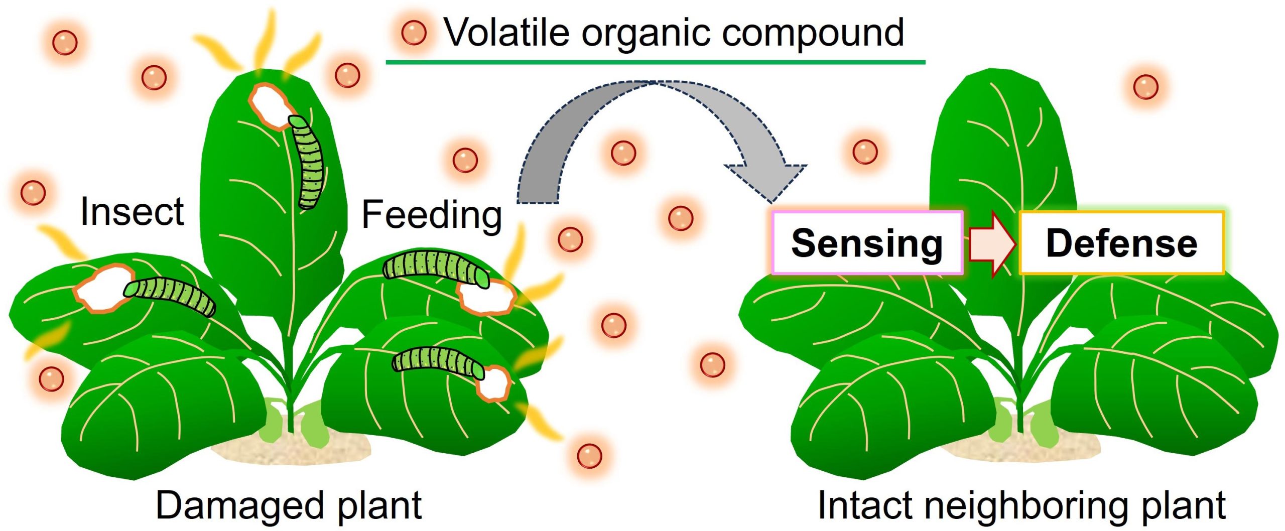
Plants emit volatile organic compounds (VOCs) into the atmosphere upon mechanical damage or insect attacks. Undamaged neighboring plants sense the released VOCs as danger cues to activate defense responses against upcoming threats (Figure 1). This phenomenon of airborne communication among plants through VOCs was first documented in 1983 and has since been observed in more than 30 different plant species. However, the molecular mechanisms underlying VOC perception to defense induction remain unclear.
Groundbreaking Visualization of Plant Conversations
The team, led by Professor Masatsugu Toyota (Saitama University, Japan), visualized plant-plant communications via VOCs in real-time and revealed how VOCs are taken up by plants, initiating Ca2+-dependent defense responses against future threats.
This groundbreaking research will be published in the journal Nature Communications on October 17, 2023. Yuri Aratani and Takuya Uemura led the work as a Ph.D. student and a postdoctoral researcher, respectively, in Toyota’s lab and collaborated with Professor Kenji Matsui at Yamaguchi University, Japan.
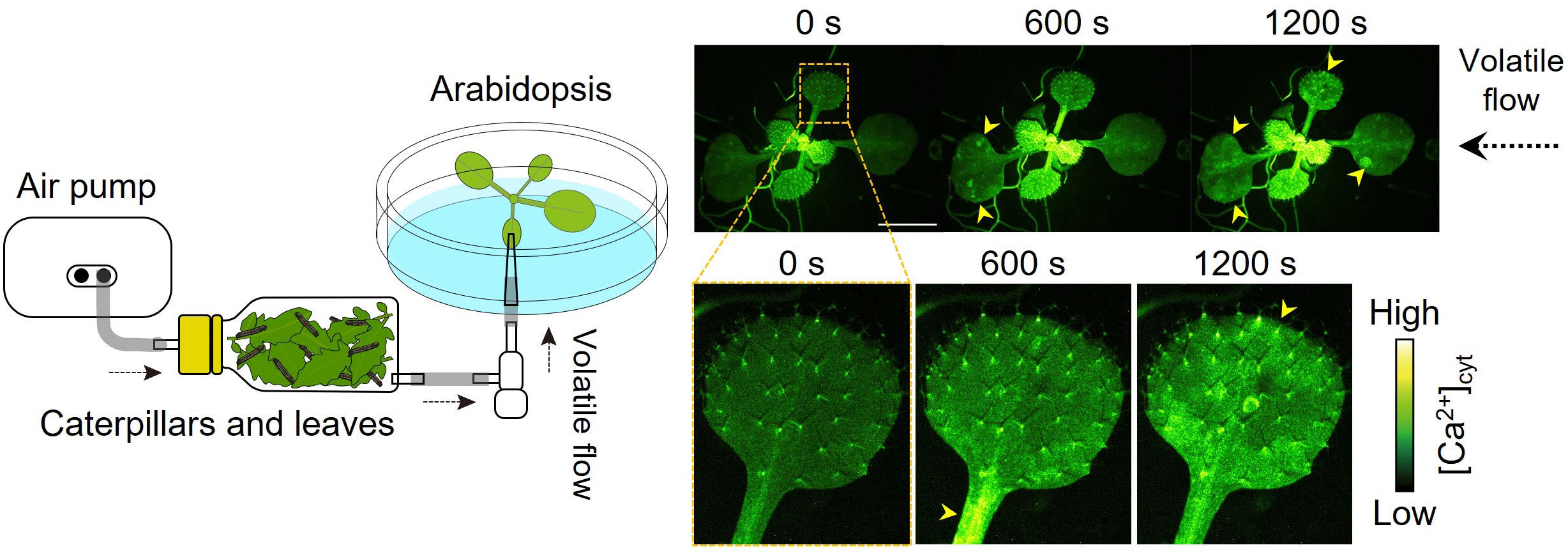
Video 1: Ca2+ signals were induced by VOCs released from insect-damaged plants (arrows). Credit: Masatsugu Toyota/Saitama University
“We constructed equipment to pump VOCs emitted from plants fed by caterpillars onto undamaged neighboring plants and combined it with a wild-field, real-time fluorescent imaging system,” says Toyota. This innovative setup visualized bursts of fluorescence spreading in a mustard plant Arabidopsis thaliana after exposure to VOCs emitted from the insect-damaged plants (Figure 2; Video 1). The plants create fluorescent protein sensors for intracellular Ca2+ and therefore, changes in intracellular Ca2+ concentration can be monitored by observing changes in fluorescence.
“In addition to insect attacks, VOCs released from manually smashed leaves induced Ca2+ signals in undamaged neighboring plants,” says Toyota (Video 2).
Identification of Key VOCs and Their Impact
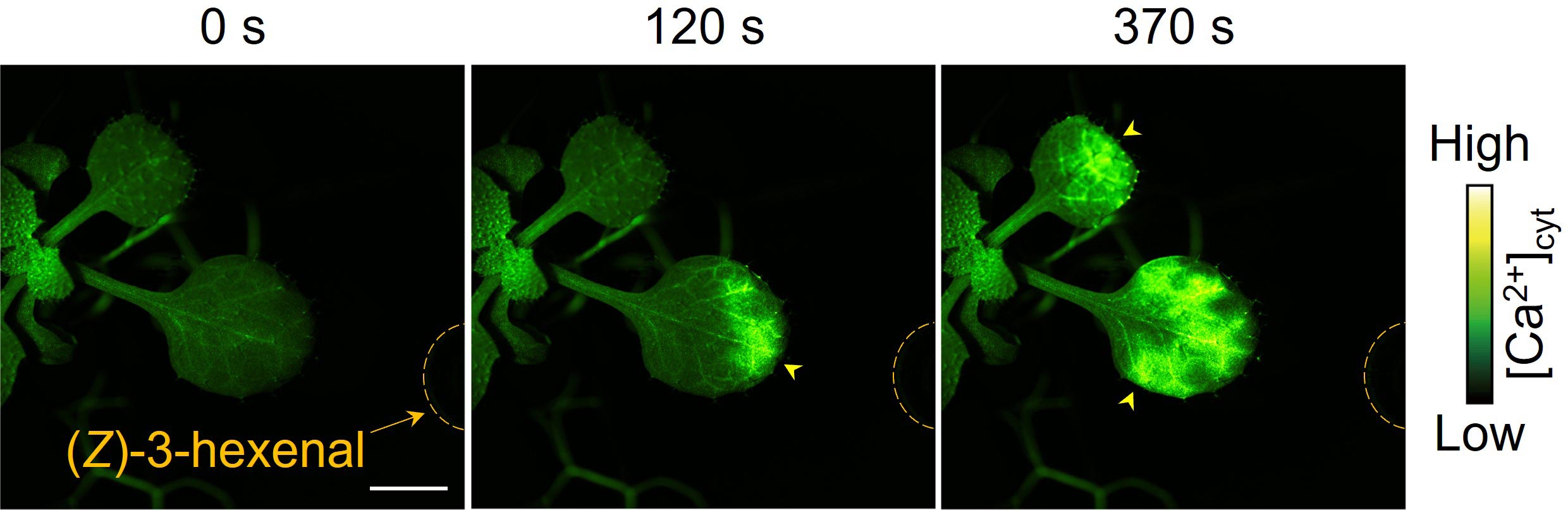
To identify what type of VOCs induced Ca2+ signals in plants, Toyota’s team of scientists investigated various VOCs known to induce defense responses in plants. They found that two VOCs, (Z)-3-hexenal (Z-3-HAL) and (E)-2-hexenal (E-2-HAL), both six-carbon aldehydes, induce Ca2+ signals in Arabidopsis (Figure 3; Video 3). Z-3-HAL and E-2-HAL are airborne chemicals with grassy smells and are known as green leaf volatiles (GLVs) emitted from mechanically- and herbivore-damaged plants.
Video 2: Ca2+ signals were induced by VOCs released from manually smashed plants. Credit: Masatsugu Toyota/Saitama University
Exposing Arabidopsis to Z-3-HAL and E-2-HAL resulted in the upregulation of defense-related genes. To understand the relationship between the Ca2+ signals and the defense responses, they treated Arabidopsis with the Ca2+ channel inhibitor, LaCl3 and the Ca2+ chelating agent, EGTA. These chemicals suppressed both the Ca2+ signals and the induction of defense-related genes, providing evidence that Arabidopsis perceives GLVs and activates defense responses in a Ca2+-dependent manner.
Guard Cells: Plants’ Gateway to Awareness
They also identified which specific cells exhibited the Ca2+ signals in response to GLVs by engineering transgenic plants expressing the fluorescent protein sensors exclusively in guard, mesophyll, or epidermal cells. Upon Z-3-HAL exposure, Ca2+ signals were generated in guard cells within approximately 1 minute and then in mesophyll cells, whereas epidermal cells generated Ca2+ signals more slowly (Video 4). Guard cells are bean-shaped cells on plant surfaces and form stomata, small pores that connect inner tissues and the atmosphere.
Video 3: Airborne Z-3-HAL (in the tube on the right side) induced Ca2+ signals in Arabidopsis leaves. Credit: Masatsugu Toyota/Saitama University
“Plants do not possess a “nose,” but stomata serve as a plant gateway mediating rapid GLV entry into interspaces in leaf tissues,” says Toyota. In fact, they found that pretreating with abscisic acid (ABA), one of the phytohormones known for its ability to close stomata, reduced Ca2+ responses in wild-type leaves. On the other hand, mutants with impaired ABA-induced stomatal closures maintained normal Ca2+ signals in leaves even when treated with ABA.
“We have finally unveiled the intricate story of when, where, and how plants respond to airborne ‘warning messages’ from their threatened neighbors,” he says. “This ethereal communication network, hidden from our view, plays a pivotal role in safeguarding neighboring plants from imminent threats in a timely manner,” he adds.
Video 4: Airborne Z-3-HAL induced Ca2+ signals in guard (left video), mesophyll (central video), and then epidermal cells (right video) in Arabidopsis leaves. Credit: Masatsugu Toyota/Saitama University
This pioneering research not only deepens our appreciation for the astonishing world of plants but also underscores the remarkable ways in which nature has equipped them to thrive and adapt in the face of adversity. The profound implications of these findings resonate far beyond the boundaries of plant science, offering a glimpse into the intricate tapestry of life on Earth.
Reference: “Green leaf volatile sensory calcium transduction in Arabidopsis” by Yuri Aratani, Takuya Uemura, Takuma Hagihara, Kenji Matsui and Masatsugu Toyota, 17 October 2023, Nature Communications.
DOI: 10.1038/s41467-023-41589-9
Funding: Japan Society for the Promotion of Science, Japan Science and Technology Agency, Shiraishi Foundation of Science Development
Never miss a breakthrough: Join the SciTechDaily newsletter.
Follow us on Google and Google News.