Brain activity patterns stem from stationary resonant waves, as revealed by ultrafast MRI in rats, suggesting new insights into brain communication and disorder diagnostics.
It’s been over 20 years since neuroimaging studies – using functional magnetic resonance imaging (fMRI), a widely-used technology to capture live videos of brain activity – have been detecting brain-wide complex patterns of correlated brain activity that appear disrupted in a wide range of neurological and psychiatric disorders. These patterns form spontaneously, even at rest when no particular task is being performed, and have been detected not only in humans but also across mammals, including monkeys and rodents.
Although such spatial patterns of correlated activation have been consistently detected across neuroimaging centers around the world, the nature of these correlations was not clear. “We do not yet fully understand how the brain communicates over long distances. We know that distant areas exhibit signal correlations, and that they are implicated in brain function, but we do not completely understand their nature,” says Noam Shemesh, principal investigator of the Preclinical MRI Lab at the Champalimaud Foundation, in Lisbon, and senior author of a study published on February 6th, 2023, in the journal Nature Communications.
“In this study, we wanted to understand what lies underneath those correlations and investigate the mechanisms involved,” stresses Shemesh.
Resonance Hypothesis and Oscillatory Patterns
A number of theoretical works had proposed that these patterns could be explained by standing waves (whose peaks and troughs do not move in space) resonating in the brain structure – that is, by waves analogous to the modes of vibration in musical instruments. But there was little experimental evidence to support this hypothesis due to the poor temporal resolution of fMRI, reaching only an image or two per second. “If we could find that the spatial patterns oscillate, this would provide evidence supporting the resonance hypothesis,” says Joana Cabral, first author of the study, from the Life and Health Sciences Research Institute of the University of Minho and a visiting scientist in Shemesh’s lab since 2019.
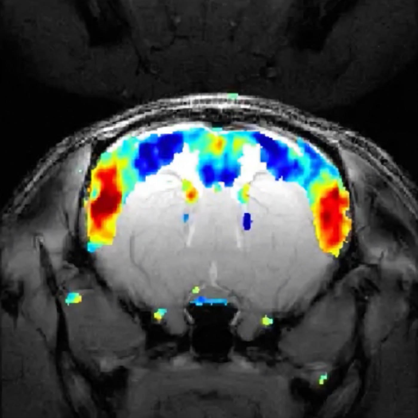
The video shows that brain activity captured with fMRI can be reconstructed as the superposition of a small number of macroscopic stationary waves, or resonant modes, oscillating in time. Credit: Joana Cabral
So what the team did was to speed up image acquisition, and they discovered that the signals in distant brain regions actually oscillate together in time. “These oscillatory patterns look like a higher-dimensional analogue of resonance modes in musical instruments; they are akin to reverberations, to echoes inside the brain,” says Cabral.
“Our data show that the complex spatial patterns are a result of transiently and independently oscillating underlying modes, just like individual instruments participate in creating a more complex piece in an orchestra,” says Shemesh. “The distinct modes, each contributing something to the overall picture at different time scales and different wavelengths, can be added up together, generating complex macroscopic patterns similar to the ones observed experimentally [see below]. To our knowledge, this is the first time that brain activity captured with fMRI is reconstructed as the superposition of standing waves,” he points out.
The new study thus strongly points to a key role for these resonant waves, or modes, in brain function. These resonant phenomena, the authors believe, are at the root of the coherent, coordinated brain activity that is needed for normal brain function as a whole.
Ultrafast MRI
The researchers detected the resonant modes in rats in the resting state, which means the animals were not subjected to any specific external stimulus. Indeed, no tasks were needed, for as already mentioned, even when we (and mammals in general) are doing nothing in particular, our brains continue to generate spontaneous activity patterns that can be captured by fMRI.
To visualize the oscillations, the researchers created “videos” of activity using the potent ultrahigh-field experimental MRI scanner in Shemesh’s lab and performed ultrafast experiments developed some time ago by that lab for other purposes.
“Noam and I met in 2019, and we decided to obtain recordings of brain activity at the maximum temporal resolution we could achieve in the 9.4 Tesla scanner at his lab,” recalls Cabral. “Noam designed the experiments, Francisca Fernandes [the third author of the study] performed them, and I did the data analysis and the visualization. Noam managed to achieve a temporal resolution of 26 images per second, and thus obtained 16,000 images per 10-minute scan (instead of 600 images at the typical resolution of one image per second).”
Like waves in the ocean
“When we first saw the videos of the recorded brain activity, we saw clear waves of activity, like waves in the ocean, propagating in complex patterns within the cortex and the striatum [a subcortical region of the forebrain],” says Cabral. “And we found that the signals could be described by the superposition of a small number of macroscopic stationary waves, or resonant modes, oscillating in time. Notably, each standing wave was found to cover extended areas of the brain, with peaks distributed in distinct cortical and subcortical structures, forming functional networks.”
The researchers experimented with rats in three different conditions: sedated, lightly anesthetized, and deeply anesthetized. (In fact, the animals were lightly sedated in the resting state, to avoid any discomfort to them.) “The spatial configuration of these stationary waves was very consistent across rats scanned in the same condition,” Cabral points out.
Shemesh adds: “We showed that brain functional networks are driven by resonance phenomena. This explains the correlations that are otherwise observed when you do slow imaging. Long-range brain interactions are governed by a ‘flow’ of information that is oscillatory and repetitive.”
Pathological states
They also found that increasing the amount of anesthetic reduces the number, frequency, and duration of the resonant stationary waves. As already mentioned, previous studies have shown that certain patterns of brain activation are consistently altered in disorders of consciousness. So this experimental design, says Cabral, was actually also meant to mimic different pathological states.
“Functional networks appear disrupted in several neurological and psychiatric disorders” she points out. If confirmed in humans, she speculates, their results could also lead to the use of resonant modes as biomarkers for disease.
“Our study also provides a new ‘lead’ in looking at disease,” corroborates Shemesh. “We know that long-range brain activity is strongly impacted in disease, but we do not understand why or how. Understanding the mechanism of long-range interactions could lead to a completely new way of characterizing disease and hinting on the type of treatment that may be necessary: for example, if a specific resonant mode was missing from a patient, we might want to find ways to stimulate that particular mode.”
More work will obviously be needed to confirm all these results, the researchers agree, and whether they are replicable in humans. But “once we understand better the nature of functional networks, we can design informed strategies to modulate these network patterns,” says Cabral.
This is precisely the subject of the researchers’ new project, “BRAINSTIM: Predicting stimulation strategies to modulate interactions between brain areas.” Funded by the “la Caixa” Foundation and the Portuguese bank BPI, with 300,000 euros, it is a collaboration between the Life and Health Sciences Institute of the University of Minho and the Champalimaud Foundation – and its aim is to better understand the impact of distinct pharmacological and electromagnetic brain stimulations in the modulation of these macroscale oscillatory modes.
Reference: “Intrinsic macroscale oscillatory modes driving long-range functional connectivity in female rat brains detected by ultrafast fMRI” by Joana Cabral, Francisca F. Fernandes and Noam Shemesh, 6 February 2023, Nature Communications.
DOI: 10.1038/s41467-023-36025-x
Never miss a breakthrough: Join the SciTechDaily newsletter.
Follow us on Google and Google News.