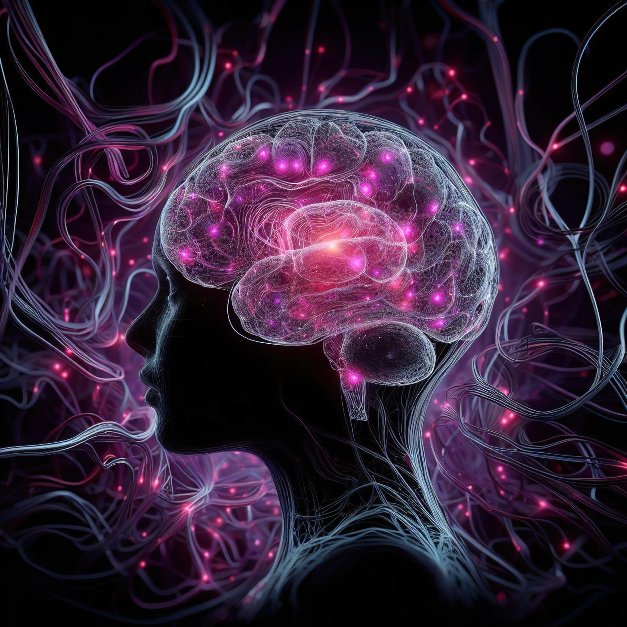
Researchers have identified a brain biomarker, the “somato-visual” marker, using MRI scans. It shows distinct connectivity patterns in psychosis patients and could allow for earlier diagnosis and intervention, transforming care and treatment strategies.
The standard approach to diagnosing psychosis relies on a diagnostic interview, but what if it were possible to identify the condition before the first symptoms appeared? Researchers at the Del Monte Institute for Neuroscience at the University of Rochester are exploring a potential brain biomarker that could pave the way for earlier interventions and more personalized treatment.
“Establishing such biomarkers could provide a key step in changing how we care for, treat, and offer interventions to people with psychosis,” said Brian Keane, PhD, assistant professor of Psychiatry, Center for Visual Science, and Neuroscience at the University of Rochester Medical Center. Keane recently co-authored an article in Molecular Psychiatry that identifies how MRI scans could reveal brain differences in people with psychosis. “Aside from potentially predicting future psychosis onset, biomarkers could also help stratify patients into clinically meaningful subgroups and suggest new options for treatment or intervention.”
Brain Connectivity and Biomarkers for Psychosis
Using data collected by the Human Connectome Early Psychosis Project, researchers looked at MRI scans from 159 participants. These included 105 who developed a psychotic disorder up to five years prior to testing. In the brains of participants with psychosis, researchers found that sensory regions in the cortex were more weakly connected to each other and more strongly connected to the thalamus, the brain’s information relay station.
These differences were confined to the somatomotor network, which processes bodily movement and sensations, and a visual network, which generates representations of objects, faces, and complex features. Combining the dysconnectivity patterns across these two networks allowed the researchers to create a “somato-visual” biomarker.
Unique Insights into Brain Dysconnectivity
Previous research has suggested that abnormal brain connectivity exists prominently in the sensory networks of people with schizophrenia, but it remained unclear which networks were most responsible or whether dysconnectivity could be explained by other illness factors, such as antipsychotic use, anxiety, or stress.
“What makes this biomarker unique is its large effect size, its robustness to over a dozen common confounds, and its high reliability across multiple scans. A single five-minute scan could potentially improve our ability to predict which at-risk individuals will transition to a psychotic disorder, which in turn could allow for more timely treatments or interventions,” Keane said. “It also gives us a place to keep looking. An important next step will be to determine if the somato-visual biomarker emerges before or as psychosis begins.”
Reference: “Functional dysconnectivity of visual and somatomotor networks yields a simple and robust biomarker for psychosis” by Brian P. Keane, Yonatan T. Abrham, Michael W. Cole, Brent A. Johnson, Boyang Hu and Carrisa V. Cocuzza, 4 October 2024, Molecular Psychiatry.
DOI: 10.1038/s41380-024-02767-3
Additional authors include Yonatan Abrham, Boyang Hu, and Brent Johnson of the University of Rochester, Carrisa Cocuzza of Yale University, and Michael Cole of Rutgers University. This work was supported by a K01 grant and a Psychiatry Department pilot grant at the University of Rochester.
Never miss a breakthrough: Join the SciTechDaily newsletter.
Follow us on Google and Google News.