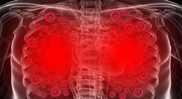
Scientists have unveiled the initial map detailing how human lung cells respond on a molecular level to SARS-CoV-2 infection. They’ve pinpointed specific host proteins and pathways in lung cells that undergo alterations upon infection, offering valuable insights into the disease’s progression and potential new targets for COVID-19 treatment.
Researchers identify clinically approved medications that can be re-purposed for COVID-19 treatment.
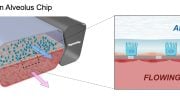
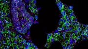
In a multi-group collaborative involving the National Emerging Infectious Disease Laboratories (NEIDL), the Center for Regenerative Medicine (CReM), and the Center for Network Systems Biology (CNSB), scientists have reported the first map of the molecular responses of human lung cells to infection by SARS-CoV-2. By combining bioengineered human alveolar cells with sophisticated, highly precise mass spectrometry technology, Boston University School of Medicine (BUSM) researchers have identified host proteins and pathways in lung cells whose levels change upon infection by the SARS-CoV-2, providing insights into disease pathology and new therapeutic targets to block COVID-19.
They found a crucial type of protein modification called “phosphorylation” becomes aberrant in these infected lung cells. Phosphorylation of proteins play a major role in regulating protein function inside the cells of an organism and both protein abundance and protein phosphorylation are typically highly controlled processes in the case of normal/healthy cells. However, they discovered that SARS-CoV-2 throws the lung cells into disarray, causing abnormal changes in protein amounts and frequency of protein phosphorylation inside these cells. These abnormal changes help the virus to multiply eventually destroy the cells. The destruction of infected cells may result in widespread lung injury.
According to the researchers, as soon as the SARS-CoV-2 enters the lung cells, it rapidly begins to exploit the cell’s core resources, which are otherwise required for the cell’s normal growth and function. “The virus uses these resources to proliferate while evading attack by the body’s immune system. In this way new viruses form which subsequently exit the exhausted and brutally damaged lung cell, leaving them to self-destruct. These new viruses then infect other cells, where the same cycle is repeated,” explains corresponding author Andrew Emili, PhD, professor of biochemistry at BUSM.
The researchers examined lung alveolar cells from one to 24 hours after infection with SARS-CoV-2 to understand what changes occur in lung cells immediately (at one, three and six hours after infection by SARS-CoV-2) and what changes occur later (at 24 hours after infection). These changes were then compared to uninfected cells. All proteins from infected and uninfected alveolar cells, corresponding to the different time-points were extracted and labeled with unique barcoding tags called “tandem mass tag.” These tags, which can be accurately detected only by a mass spectrometer, permit robust quantification of protein and phosphorylation abundance in cells.
“Our results showed that in comparison to normal/uninfected lung cells, SARS-CoV-2 infected lung cells showed dramatic changes in the abundance of thousands of proteins and phosphorylation events,” said Darrell Kotton, MD, professor of pathology & laboratory medicine at BUSM and director of the CReM.
“Moreover, our data also showed that the SARS-CoV-2 virus induces a significant number of these changes as early as one hour post infection and lays the foundation for a complete hijack of the host lung cells,” adds Elke Mühlberger, PhD, associate professor of microbiology and principal investigator at the NEIDL.
“There are important biological features specific to lung cells that are not reproduced by other cell types commonly used to study viral infection,” said Andrew Wilson, MD, associate professor of medicine at BUSM and CReM investigator. “Studying the virus in the context of the cell type that is most damaged in patients is likely to yield insights that we wouldn’t be able to see in other model systems.”
The researchers also analyzed their data to identify prospective opportunities for COVID-19 treatment and found that at least 18 pre-existing clinically approved drugs (developed originally for other medical conditions/diseases) can be potentially re-purposed for use towards COVID-19 therapy. These drugs have shown exceptional promise to block the proliferation of the SARS-CoV-2 in lung cells.
The researchers believe this information is invaluable and paves the way for newer, potentially promising and more importantly, a cost-effective and time-saving therapeutic strategy to combat COVID-19.
Researchers Raghuveera Kumar Goel, PhD; Adam Hume, PhD; Jessie Huang, PhD; Kristy Abo, BA; Rhiannon Werder, PhD and Ellen Suder, BS, also contributed to these findings.
These findings appear online in the journal Molecular Cell.
Reference: “Actionable Cytopathogenic Host Responses of Human Alveolar Type 2 Cells to SARS-CoV-2” by Ryan M. Hekman, Adam J. Hume, Raghuveera Kumar Goel, Kristine M. Abo, Jessie Huang, Benjamin C. Blum, Rhiannon Bree Werder, Ellen L. Suder, Indranil Paul, Sadhna Phanse, Ahmed Youssef, Kostantinos D. Alysandratos, Dzmitry Padhorny, Sandeep Ojha, Alexandra Mora-Martin, Dmitry Kretov, Peter Ash, Mamta Varma, Jian Zhao, J.J. Patten, Carlos Villacorta-Martin, Dante Bolzan, Carlos Perea-Resa, Esther Bullitt, Anne Hinds, Andrew Tilston-Lunel, Xaralabos Varelas, Shaghayegh Farhangmehr, Ulrich Braunschweig, Julian H. Kwan, Mark McComb, Avik Basu, Mohsan Saeed, Valentina Perissi, Eric J. Burks, Matthew D. Layne, John H. Connor, Robert Davey, Ji-Xin Cheng, Benjamin L. Wolozin, Benjamin J. Blencowe, Stefan Wuchty, Shawn M. Lyons, Dima Kozakov, Daniel Cifuentes, Michael Blower, Darrell N. Kotton, Andrew A. Wilson, Elke Mühlberger and Andrew Emili, 19 November 2020, Molecular Cell.
DOI: 10.1016/j.molcel.2020.11.028
Funding was provided by NIH F30HL147426 to K.M.A.; CJ Martin Fellowship from the Australian NHMRC to R.B.W.; IM Rosenzweig Award, Pulmonary Fibrosis Foundation to K.D.A.; NIH AG056318, AG064932, AG061706, and AG050471 to B.L.W.; NIH RM1 GM135136 and R21GM127952 and NSF AF1816314 to D. Kozakov; Evergrande MassCPR, C3.ai Digital Transformation Institute Award, and NIH R01HL128172, R01HL095993, R01HL122442, U01HL134745, and U01HL134766 to D.N.K.; CIHR, COVID-19 Action Initiative to B.J.B.; NIH HL007035T32 to E.L.S.; NIH R01AI125453, P01AI120943, and R01AI128364 and Massachusetts Consortium on Pathogen Readiness to J.J.P. and R.D.; NIH U01TR001810, UL1TR001430, R01DK101501, and R01DK117940 to A.A.W.; Fast Grants, Evergrande MassCPR, and NIH R01AI133486, R21AI135912, R21AI137793, and R21AI147285 to E.M.; and NIH 1UL1TR001430, RO1AG064932, and RO1AG061706 to A.E.
Disclosures: B.L.W. declares a position as CSO of Aquinnah Pharmaceuticals. A.E. and D.N.K. declare industry funding from Johnson & Johnson, Merck, and Novartis.

But it has a 99% survival rate.
That is some really great news. I just wish these professionals who are working on understanding and combating this disease on a cellular level got more credit for all of the hours of laborious struggle they’ve endured to get to this point so quickly.