A new study introduces a novel approach to predicting the course of uveitis through retinal blood flow analysis, potentially transforming patient monitoring and clinical trial outcomes.
An estimated five to ten percent of blindness worldwide is due to the rare inflammatory eye disease uveitis. Intermediate uveitis is often associated with a chronic course of the disease and the need for immunosuppressive therapy. Intermediate uveitis primarily causes inflammation of the vitreous body, but blood flow to the retina can also be restricted.
Researchers at the Eye Clinic of the University Hospital Bonn (UKB) and the University of Bonn have tested optical coherence tomography angiography as a new imaging monitoring method. The blood flow in the retinal vessels is associated with the severity of inflammation and allows conclusions to be drawn about the future course of the disease.
Accordingly, this method could be used to monitor the disease and identify patients at risk of a future worsening of the disease. The results have now been published in Nature Scientific Reports.
The Challenge of Intermediate Uveitis
Blurred vision and streaks in front of the eye — those affected by the rare disease intermediate uveitis have no pain.
“But the consequences can be serious: around five to ten percent of blindness worldwide is caused by uveitis. It is a rare disease and intermediate uveitis in particular is often associated with a long course of the disease and the need for immunosuppressive therapy,” says Dr. Maximilian Wintergerst from the UKB Eye Clinic, who also conducts research at the University of Bonn. There are different forms of the disease.
In the case of intermediate uveitis, it is primarily the vitreous body in the eye that becomes inflamed. This is the gelatinous mass that fills the eye. However, the retinal vessels can also be inflamed, as the research group at the UKB and the University of Bonn, among others, was able to show in preliminary work.
Importance of Early Detection
“It is important to recognize an increase in inflammatory activity in good time,” says Wintergerst. This allows treatment to be adjusted if necessary, which can preserve visual acuity and prevent further complications. However, there are currently only a few objective parameters that can be used to reliably detect a worsening of the disease.
Most criteria for assessing disease activity are based on clinical examination and are comparatively subjective and not always reliable. Therefore, researchers from the UKB Eye Clinic together with colleagues from Medical Biometry at the University Hospital Bonn investigated new high-resolution imaging-based methods to determine disease activity and complications in uveitis.
“Objective markers of inflammatory activity could not only improve monitoring in everyday clinical practice, but would also provide additional quantitative endpoints for future randomized clinical trials,” adds Wintergerst.

Innovations in Monitoring Disease Progression
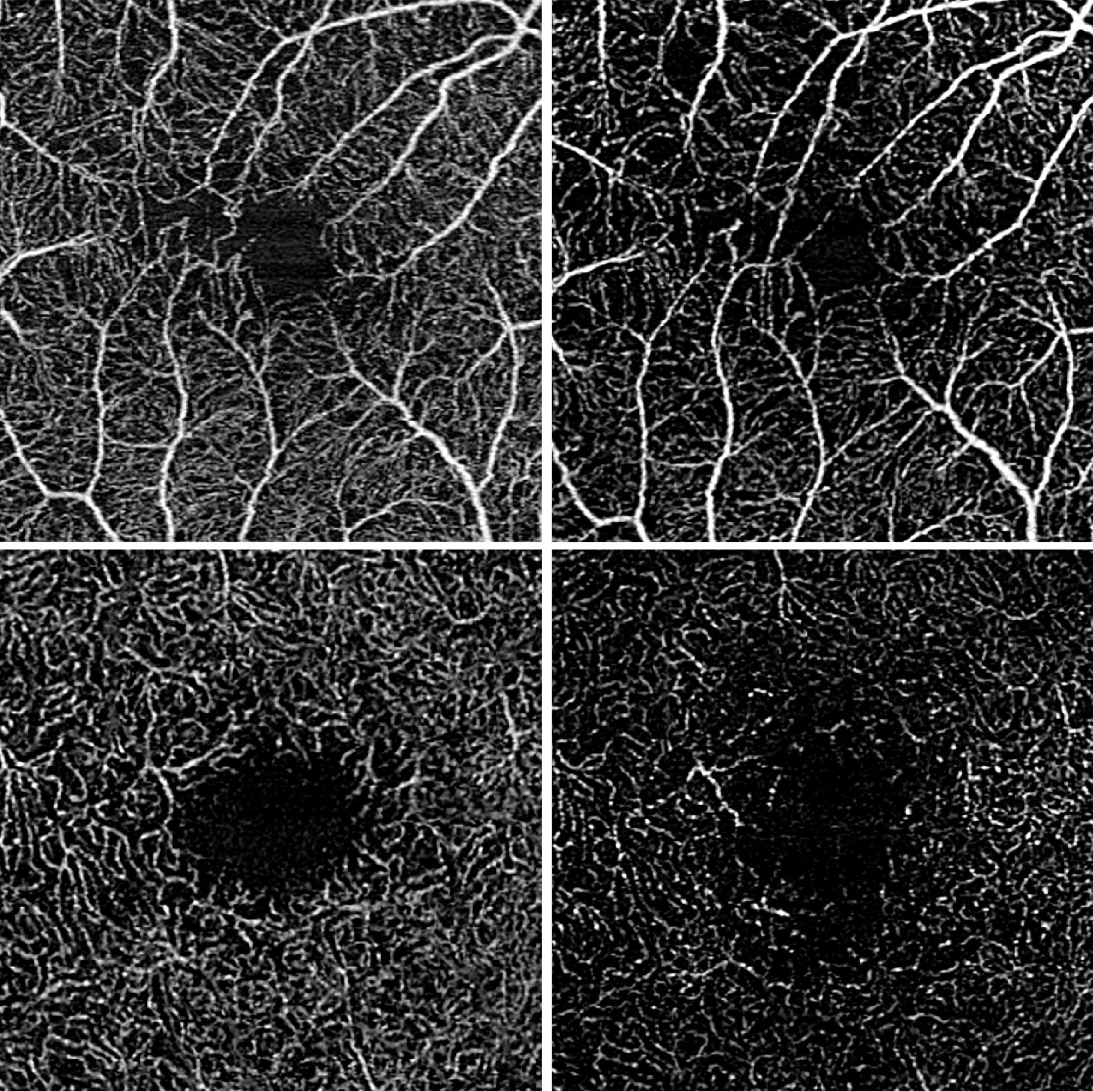
“Optical coherence tomography angiography enables non-contact, non-invasive examination of the retina and the underlying choroid. The retina is scanned successively using harmless, weak laser light, which allows tomographic images of the individual retinal layers to be generated,” explains Wintergerst.
By comparing several images taken in quick succession, the blood flow can be detected, which allows conclusions to be drawn about the blood supply to the retinal vessels.
The researchers from the UKB and the University of Bonn then calculated the blood flow density of the central retina and analyzed how this differs between eyes with stable disease, eyes with an increase in disease activity and eyes with a decrease in disease activity.
The researchers examined a total of 52 study participants and were able to show that the blood flow density differed between the three groups examined. An increase in disease activity was associated with a decrease in blood flow density, while a decrease in disease activity was associated with an increase in blood flow density.
Predictive Power of Blood Flow Density
In addition, the Bonn researchers used a statistical model, which included over 300 eye examinations, to investigate the predictive power of current blood flow density for the future course of the disease. This showed that a reduced blood flow density was significantly associated with a future deterioration in central visual acuity.
“In the future, the data obtained could enable us to identify patients with a high risk of disease progression at an earlier stage, for example in order to monitor them particularly closely,” says Prof. Dr. Dr. Robert Finger, co-author of the study and now Director of the Eye Clinic at the University Medicine Mannheim (UMM).
“We could use this parameter as an endpoint in future randomized clinical trials in order to potentially generate better evidence for the treatment of this rare disease.”
Conclusion and Future Implications
“In the current study, we show how objective parameters for disease activity in uveitis can be determined using high-resolution, digital non-invasive imaging,” says Prof. Dr. Frank Holz, Director of the UKB Eye Clinic. “This is an important prerequisite for improving the monitoring of uveitis in the future.”
Reference: “Vessel density on optical coherence tomography angiography is prognostic for future disease course in intermediate uveitis” by Maximilian W. M. Wintergerst, Nicholas R. Merten, Moritz Berger, Jan H. Terheyden, Lennart J. Overbeck, Matthias Schmid, Frank G. Holz and Robert P. Finger, 5 February 2024, Scientific Reports.
DOI: 10.1038/s41598-023-49926-0
Never miss a breakthrough: Join the SciTechDaily newsletter.
Follow us on Google and Google News.