Scientists have mapped the yellow fever virus in unprecedented detail.
Researchers at the University of Queensland have produced the first high-resolution images of the yellow fever virus (YFV), a serious mosquito-borne pathogen that attacks the liver and can cause fever, jaundice, internal bleeding, and organ failure in severe cases.
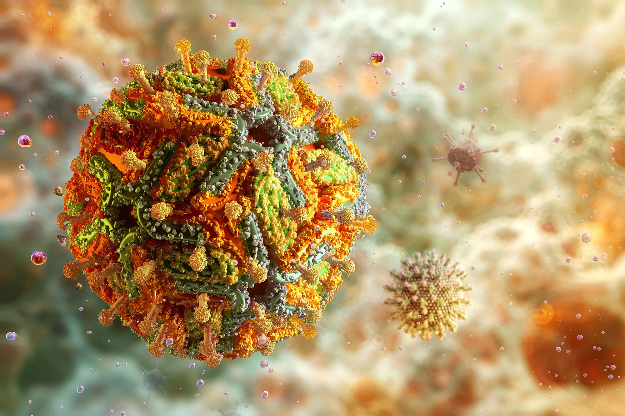
Yellow fever belongs to the Flavivirus genus, a group that also includes dengue, Zika, and West Nile viruses. It is composed of a spherical particle containing a single strand of RNA encased within a protective protein shell and lipid membrane.
Using advanced imaging methods, the research team identified distinct structural variations between the vaccine strain (YFV-17D) and the more virulent, disease-causing versions of the virus.
Dr. Summa Bibby from UQ’s School of Chemistry and Molecular Bioscience explained that, although scientists have studied yellow fever for decades, this marks the first time a complete three-dimensional model of a fully mature yellow fever virus particle has been visualized at near-atomic resolution.
“By utilizing the well-established Binjari virus platform developed here at UQ, we combined yellow fever’s structural genes with the backbone of the harmless Binjari virus and produced virus particles that could be safely examined with a cryo-electron microscope,” Dr. Bibby said. “The particles of the vaccine strain had a smooth and stable surface layer, while the particles of the virulent strain had bumpy, uneven surfaces.”
Distinct Structural Differences Between Strains
The differences change how the body’s immune system recognizes the virus.
“The bumpier, irregular surface of the virulent strains exposes parts of the virus that are normally hidden, allowing certain antibodies to attach more easily,” Dr. Bibby said.
“The smooth vaccine particles keep those regions covered, making them harder for particular antibodies to reach.”
Yellow fever is a major public health concern in parts of South America and Africa and with no approved antiviral treatments, vaccination is the primary means of prevention.
Professor Daniel Watterson said the discovery provides crucial new insights into yellow fever biology and opens the door to improved vaccine design and antiviral strategies for it and other orthoflaviviruses.
“The yellow fever vaccine remains effective against modern strains and seeing the virus in such fine detail lets us better understand why the vaccine strain behaves the way it does,” Professor Watterson.
“We can now pinpoint the structural features that make the current vaccine safe and effective.
“The findings could even inform future vaccine design for related viruses like dengue, Zika, and West Nile.”
Reference: “A single residue in the yellow fever virus envelope protein modulates virion architecture and antigenicity” by Summa Bibby, James Jung, Yu Shang Low, Alberto A. Amarilla, Natalee D. Newton, Connor A. P. Scott, Jessica Balk, Yi Tian Ting, Morgan E. Freney, Benjamin Liang, Timothy Grant, Fasséli Coulibaly, Paul Young, Roy A. Hall, Jody Hobson-Peters, Naphak Modhiran and Daniel Watterson, 26 September 2025, Nature Communications.
DOI: 10.1038/s41467-025-63038-5
Funding: National Health and Medical Research Council
Never miss a breakthrough: Join the SciTechDaily newsletter.
Follow us on Google and Google News.