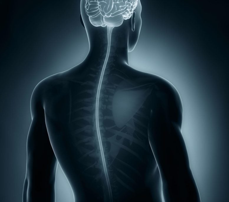
A hybrid biomaterial that has been successfully synthesized could be utilized to treat spinal injuries.
A unique material developed at the University of Limerick in Ireland has demonstrated considerable potential in the treatment of spinal cord injuries.
Exciting new research conducted at the Bernal Institute at the University of Limerick (UL) and published in the journal Biomaterials Research, has made significant strides in the area of spinal cord tissue repair.
New hybrid biomaterials developed at UL in the form of nanoparticles and building on existing practice in the tissue engineering field, were successfully synthesized to promote repair and regeneration following spinal cord injury, according to the researchers.
The UL team led by Professor Maurice N Collins, Associate Professor, School of Engineering at UL, and lead author Aleksandra Serafin, a Ph.D. candidate at UL, used a new kind of scaffolding material and a unique new electrically conducting polymer composite to promote new tissue growth and generation that could advance the treatment of spinal cord injury.
Conductive Polymers in Tissue Engineering
“Spinal Cord Injury remains one of the most debilitating traumatic injuries a person can sustain during their lifetime, affecting every aspect of the person’s life,” explained Professor Collins.
“The debilitating disorder results in paralysis below the level of injury and, in the US alone, the annual healthcare costs for SCI patient care are $9.7 billion. As there is currently no widely available treatment, continuous research into this field is crucial to find a treatment to improve the patient’s quality of life, with the research field turning towards tissue engineering for novel treatment strategies.
“The field of tissue engineering aims to solve the global problem of shortages of donated organs and tissues, in which a new trend has emerged in the form of conductive biomaterials. Cells in the body are affected by electrical stimulation, especially cells of a conductive nature such as cardiac or nerve cells,” Professor Collins explained.
Overcoming Biocompatibility Challenges with Novel PEDOT Nanoparticles
The research team describes a growing interest in the use of electroconductive tissue-engineered scaffolds that have emerged due to the improved cell growth and proliferation when cells are exposed to a conductive scaffold.
“Raising the conductivity of biomaterials to develop such treatment strategies typically centers on the addition of conductive components such as carbon nanotubes or conductive polymers such as PEDOT:PSS, which is a commercially available conductive polymer that has been used to date in the tissue engineering field,” explained lead author Aleksandra Serafin, a Ph.D. candidate in the Bernal and at UL’s Faculty of Science and Engineering.
“Unfortunately, severe limitations persist when using the PEDOT:PSS polymer in biomedical applications. The polymer relies on the PSS component to allow it to be water soluble, but when this material is implanted in the body, it displays poor biocompatibility.
“This means that upon exposure to this polymer, the body has potential toxic or immunological responses, which are not ideal in an already damaged tissue which we are trying to regenerate. This severely limits which hydrogel components can be successfully incorporated to create conductive scaffolds,” she added.
Promising Results in Animal Models
Novel PEDOT nanoparticles (NPs) were developed in the study to overcome this limitation. Synthesis of conductive PEDOT NPs allows for the tailored modification of the surface of the NPs to achieve desired cell response and increase the variability of which hydrogel components can be incorporated, without the required presence of PSS for water solubility.
In this work, hybrid biomaterials comprised of gelatin and immunomodulatory hyaluronic acid, a material that Professor Collins has developed over many years at UL, were combined with the developed novel PEDOT NPs to create biocompatible electroconductive scaffolds for targeted spinal cord injury repair.
A complete study of the structure, property, and function relationships of these precisely designed scaffolds for optimized performance at the site of injury was carried out, including in-vivo research with rat spinal cord injury models, which was undertaken by Ms. Serafin during a Fulbright research exchange to the University of California San Diego Neuroscience Department, who was a partner on the project.
“The introduction of the PEDOT NPs into the biomaterial increased the conductivity of samples. In addition, the mechanical properties of implanted materials should mimic the tissue of interest in tissue-engineered strategies, with the developed PEDOT NP scaffolds matching the mechanical values of the native spinal cord,” explained the researchers.
Biological responses to the developed PEDOT NP scaffolds were studied with stem cells in-vitro and in animal models of spinal cord injury in-vivo. Excellent stem cell attachment and growth on the scaffolds were observed, they reported.
Testing showed greater axonal cell migration towards the site of spinal cord injury, into which the PEDOT NP scaffold was implanted, as well as lower levels of scarring and inflammation than in the injury model which had no scaffold, according to the study.
Overall, these results show the potential of these materials for spinal cord repair, says the research team.
Transformative Potential for Spinal Cord Injury Recovery
‘’The impact that spinal cord injury has on a patient’s life is not only physical, but also psychological, since it can severely affect the patient’s mental health, resulting in increased incidences of depression, stress, or anxiety,” explained Ms. Serafin.
“Treating spinal injuries will therefore not only allow for the patient to walk or move again but will allow them to live their lives to their full potential, which makes projects such as this one so vital to the research and medical communities. In addition, the overall societal impact of providing an effective treatment for spinal cord injuries will lead to a reduction in healthcare costs associated with treating patients.
“These results offer encouraging prospects for patients and further research into this area is planned.
“Studies have shown that the excitability threshold of motor neurons on the distal end of a spinal cord injury tends to be higher. A future project will further improve the scaffold design and create conductivity gradients in the scaffold, with the conductivity increasing towards the distal end of the lesion to further stimulate neurons to regenerate,” she added.
Reference: “Electroconductive PEDOT nanoparticle integrated scaffolds for spinal cord tissue repair” by Aleksandra Serafin, Mario Culebras Rubio, Marta Carsi, Pilar Ortiz-Serna, Maria J. Sanchis, Atul K. Garg, J. Miguel Oliveira, Jacob Koffler and Maurice N. Collins, 22 November 2022, Biomaterials Research.
DOI: 10.1186/s40824-022-00310-5
This project was funded by the Irish Research Council in partnership with Johnson & Johnson as well as the Irish Fulbright Association, which enabled a research exchange to the University of California San Diego. The faculty of Science and Engineering and the Health Research Institute at UL also provided support.
Never miss a breakthrough: Join the SciTechDaily newsletter.
Follow us on Google and Google News.
4 Comments
Question, will this only help newly injured, or will it help people who have had spinal cord for injury 20, 30 and 40 years? I have a friend who has been a paraplegic since she was around 13 years old, she has so much to give, but being locked away in a nursing home that repeatedly neglects and injures her, and that has become her prison is not acceptable.
This article actually gave me hope, for others, as I have rarely been so at my age, & psychologically stressed enough from my disability caused by a blood transfusion for an RH factor. It is “thought” that the transfusion was performed via an umbilical artery instead of a vein, then clotted; blocking nutrients from the posterior of my spine. When I was 14, Shriner’s in Chicago said I was the first in 30 years w such related “cause”.
This is so exciting and encouraging. I only pray that this new technology will not exclude me. I have waited for over 50 years for this news. I didn’t sit and wait, I raised two children on a ranch as well as worked in wonderful jobs for 25 years plus I entered the national wheelchair games in New York and received second place nationally in archery.
Please, I have a friend in Việt Nam that had a good IT job with Foxconn in Việt Nam. Last year he had a motorbike accident which caused a spinal cord injury. He lost the use of his legs. PLEASE I would love to know more about this procedure. My friend needs some good news. He lives with his parents now and tries to sell items in his hometown, it is really hard to live in Việt Nam. He would love to be able to participate in this research if possible. PLEASE let me know!! I need to know how to contact someone for more information. Thank you!!