A new DNA damage sensor allows scientists to watch repair unfold in living cells with unprecedented clarity.
Built from a natural protein domain, it binds gently and reversibly, highlighting damage without interfering with repair. The tool works in organisms as well, enabling studies of when and where DNA breaks form. Its accuracy and ease of use could boost medical research and cancer therapy development.
Relentless DNA Damage and Its Consequences
DNA inside each cell faces constant harm from sources such as sunlight, chemicals, radiation, and even the normal activities that keep our bodies functioning. Most of the time, cells repair this damage almost immediately. When those repairs do not work as they should, the resulting problems can contribute to aging, cancer, and a range of other illnesses.
Until recently, researchers struggled to watch these repair events unfold in real time. Many techniques required destroying and preserving cells at different stages, which only provided isolated snapshots rather than a full view of the process.
This footage shows the fluorescent sensors in action inside a living cell. They appear as bright green spots the moment they bind to sites of DNA damage. Credit: Richard Cardoso Da Silva
A New Live-Cell DNA Damage Sensor Emerges
Scientists at Utrecht University have now created a tool that changes this limitation. Their new DNA damage sensor makes it possible to track damage as it appears and fades inside living cells and even within living organisms. The work, published today (November 20) in Nature Communications, opens the door to research that could not be done before.
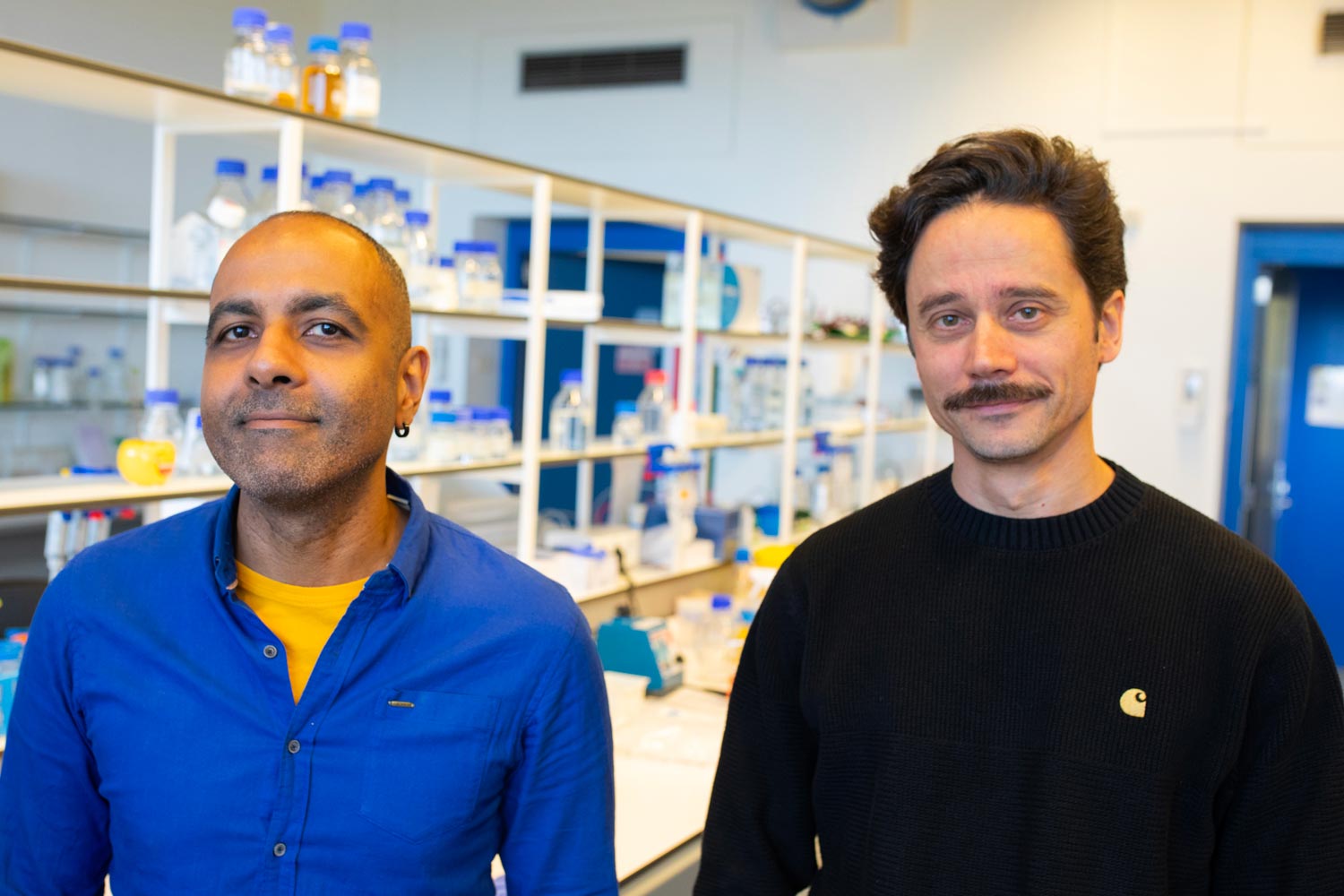
Lead researcher Tuncay Baubec describes the approach as a way to look inside a cell “without disrupting the cell.” He notes that earlier tools, including antibodies and nanobodies, tend to cling too tightly to DNA. Once they bind, they can interfere with the cell’s natural repair systems.
“Our sensor is different,” he says. “It’s built from parts taken from a natural protein that the cell already uses. It goes on and off the damage site by itself, so what we see is the genuine behavior of the cell.”
A ‘This Is Going to Work’ Moment
The tool relies on a fluorescent tag attached to a very small domain taken from one of the cell’s own proteins. This domain briefly connects to a marker that appears on damaged DNA. Because the interaction is gentle and temporary, it highlights the damaged area while leaving the cell’s repair activity undisturbed.
Biologist Richard Cardoso Da Silva, who built and evaluated the sensor, recalls when he understood its potential. “I was testing some drugs and saw the sensor lighting up exactly where commercial antibodies did,” he says. “That was the moment I thought: this is going to work.”
Capturing Continuous Repair in Real Time
The improvement over older methods is significant. Instead of running many separate experiments to capture individual moments, researchers can now track the entire sequence in a single continuous recording. They can watch damage form, see how quickly repair proteins arrive, and follow the process until the cell resolves the issue.
“You get more data, higher resolution and, importantly, a more realistic picture of what actually happens inside a living cell,” says Cardoso Da Silva.
From Lab Cells to Living Organisms
The team did not stop at cultured cells. Collaborators at Utrecht University tested the protein in the worm C. elegans, a living organism widely used in biology. The sensor performed just as well there, and revealed programmed DNA breaks that form during the worm’s development. For Baubec, this was a key moment. “It showed that the tool is not only for cells in the lab. It can be used as well in real living organisms.”
Mapping Damage and Exploring Repair Factors
The possibilities now stretch far beyond observing repair. The protein can be freely attached to other molecular parts. This means researchers can use it to map where DNA damage occurs in the genome, and also identify which proteins gather around a damaged spot. They can even move damaged DNA to different locations inside the cell nucleus to see what factors affect repair.
“Depending on your creativity and your question, you can use this tool in many ways,” says Cardoso Da Silva.
Transforming Medical and Cancer Research
Although the sensor itself is not a medical treatment, it could influence medical research. Many cancer therapies act by deliberately damaging the DNA of tumor cells. In early drug development, scientists need to measure exactly how much DNA damage a compound causes.
“Right now, clinical researchers often use antibodies to assess this,” Baubec says. “Our tool could make these tests cheaper, faster and more accurate.” The researchers also imagine uses in clinical practice, from studying natural aging processes to detecting radiation or mutagenic exposure.
Open Access for the Scientific Community
Their tool has already attracted attention. Other laboratories contacted them even before publication, eager to use the sensor in their own research on DNA repair. To support that, the team has made the tool openly available. Baubec: “Everything is online. Scientists can use it immediately.”
Reference: “Engineered chromatin readers track damaged chromatin dynamics in live cells and animals” by Richard Cardoso da Silva, Kristeli Eleftheriou, Davide C. Recchia, Vincent Portegijs, Douwe ten Bulte, Niklas Kupfer, Nathalie P. Vroegindeweij-de Wagenaar, Xabier Vergara, Ayoub Ouchene, Sander van den Heuvel and Tuncay Baubec, 20 November 2025, Nature Communications.
DOI: 10.1038/s41467-025-65706-y
Never miss a breakthrough: Join the SciTechDaily newsletter.
Follow us on Google and Google News.