
The model enables the study of the fundamental interactions that underlie neurodegenerative disease.
Researchers from Northwestern University and the University of California, Santa Barbara have engineered the first synthetic fragment of the tau protein that behaves like a prion, a type of misfolded protein known for its ability to propagate its abnormal shape to normal proteins. This synthetic “mini prion” folds into fibrils, or thread-like structures, composed of misfolded tau. These fibrils can then induce the same misfolded configuration in other healthy tau proteins.
This discovery is especially significant for understanding tauopathies, a group of neurodegenerative diseases, including Alzheimer’s disease, marked by the accumulation of misfolded tau proteins in the brain. By working with a minimal synthetic version of full-length human tau, scientists are able to more precisely replicate the structural features of pathogenic tau fibrils. This advancement may pave the way for new diagnostic tools and therapeutic strategies targeting tau-related diseases.
In the course of this research, the team also uncovered a previously unrecognized factor influencing tau misfolding: the role of water molecules near the protein surface. A disease-associated mutation, commonly used in models of tauopathies, was shown to subtly alter the structure and behavior of surrounding water. These changes, in turn, affect how readily the tau protein adopts its misfolded form.
The study was published in the Proceedings of the National Academy of Sciences.
“The scope of neurodegenerative diseases involving the protein tau is particularly broad,” said Northwestern’s Songi Han, who led the study. “It encompasses chronic traumatic encephalopathy, which is found in football players after head trauma, corticobasal degeneration, or progressive supernuclear palsy. Creating self-propagating tau fragments that can recreate the fibril structure and misfolding that is unique to each tauopathy disease is a crucial step forward in our ability to understand and model these complex diseases.”
Han is the Mark and Nancy Ratner Professor of Chemistry at Northwestern’s Weinberg College of Arts and Sciences and a member of the Chemistry of Life Processes Institute, Applied Physics Graduate Program, International Institute of Nanotechnology, Paula M. Trienens Institute for Sustainability and Energy, and Institute for Quantum Information Research and Engineering. Michael Vigers, a former Ph.D. student in Han’s laboratory, led the study and is a first author. Coauthors from UC Santa Barbara include Kenneth S. Kosik, Joan-Emma Shea and M. Scott Shell. The work also was made possible by several students and postdoctoral fellows, including Saeed Najafi, Samuel Lobo, Karen Tsay, Austin Dubose, and Andrew P. Longhini.
A chain-reaction of misfolding
In many neurodegenerative diseases, proteins misfold and clump together into harmful, highly ordered fibrils, which ultimately damage brain health but are difficult to diagnose. When a normal protein encounters the pathological tau fibrils, the normal protein changes shape to match the misfolded form. This process leads to a chain reaction, where more and more proteins transform into the misfolded, aggregation-prone state. Although this behavior is prion-like, it does not involve actual prions, which can spread contagious diseases from person to person.
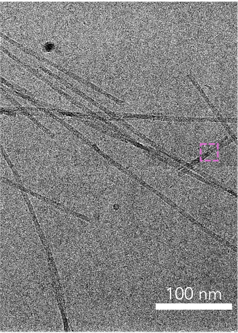
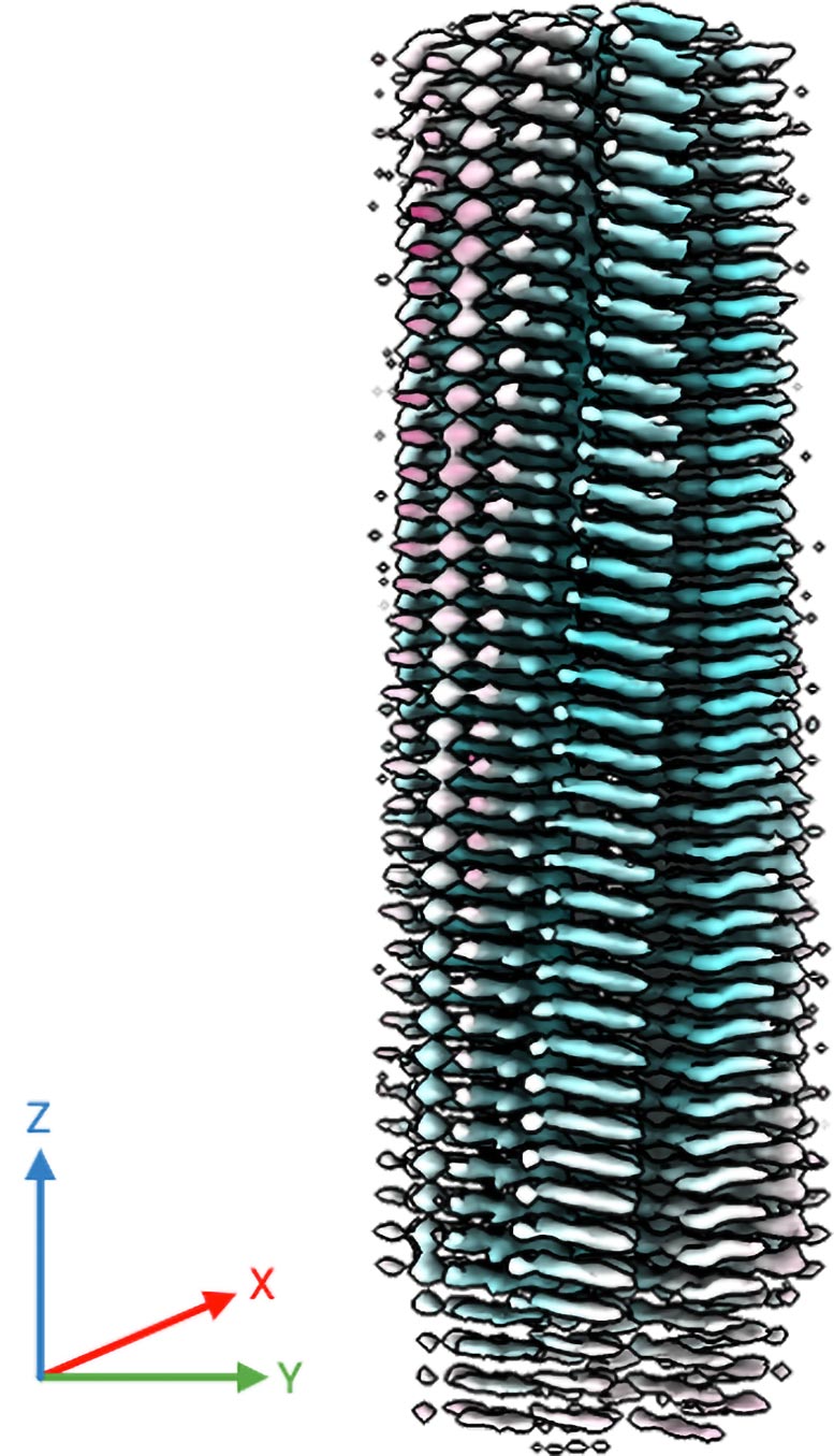
Using cryogenic electron microscopy (cryo-EM), researchers solved the structure of the fibrils from samples of brain tissue. Although pinpointing the structure was a significant breakthrough, brain samples can only be obtained after a patient dies. Despite dramatic progress and intense interest in this area, final diagnosis of tau-related neurodegenerative diseases is only possible after death.
“When people start to show signs of neurodegenerative disease, it is not diagnosed today with a biomarker,” Han said. “Physicians determine the diagnosis by administering a patient survey and by examining a collection of symptoms, like sleep patterns and memory. The bottleneck is the reliable generation of tau fibrils that recreate the critical and unique disease hallmarks to serve as targets for developing diagnostic strategies.”
A simplified model
To meet the current challenge, Han and her team sought to develop a synthetic, prion-like tau protein. Instead of recreating the entire length of the protein, which is long and unwieldy, Han’s team aimed to pinpoint the shortest piece of tau that could still adopt a misfolded shape and form disease-like fibrils.
Ultimately, Han and her team focused on a short segment of tau, dubbed jR2R3, which is just 19 amino acid segments in length. The segment contains a mutation called P301L, commonly found in many diseases. The researchers found this short peptide could form the harmful fibrils, which are the hallmark of these diseases, and act as a “seed” to template the misfolding and aggregation of full-length tau proteins.
“We made a mini version that is easier to control,” Han said. “But it does all the same things that the full-length version does. It does the seeding, causing normal tau protein to misfold and join the fibrils.”
Using cryo-EM, the team examined the structure of the synthetic fibrils. They found the P301L mutation facilitates a specific type of misfolding often observed in samples from patients with neurodegeneration. The finding suggests the mutation plays a crucial role in directing the protein to misfold.
The shape of water
Next, Han aimed to understand how the initially disordered tau proteins converge to become highly ordered fibril structures. She compared the mysterious phenomenon to throwing strands of limp spaghetti together, expecting them to form a neat stack.
“It’s impossible that an intrinsically disordered protein would just naturally fall into a perfect fold and stack that can regenerate forever,” Han said. “It doesn’t make sense.”
After hypothesizing that something must be holding the misfolded proteins together, Han found the key: water. The environment surrounding a protein, particularly the water molecules, play a crucial role in protein folding and aggregation. The P301L mutation appears to directly change the structure of the tau protein as well as change the behavior of water molecules around it.
“Water is a fluid molecule, but it still has structure,” Han said. “The mutation in the peptide might lead to a more structured arrangement of water molecules around the mutation site. This structured water influences how the peptide interacts with other molecules, pinning them together.”
In other words, organized water pins the proteins together, enabling individual strands to fold together into a neat stack. Then, using their prion-like behavior, the fibrils recruit other proteins to misfold and join the stack.
What’s next
The research team is now focused on further characterizing the properties of the synthetic, prion-like proteins. Eventually, they plan to explore potential applications, including the development of new diagnostic and therapeutic approaches for tau-related diseases.
“Once a tau fibril is formed, it doesn’t go away,” Han said. “It will grab naïve tau and fold it into the same shape. It can keep doing this forever and ever. If we can figure out how to block this activity, then we could uncover new therapeutic agents.”
Reference: “Water-directed pinning is key to tau prion formation” by Michael P. Vigers, Samuel Lobo, Saeed Najafi, Austin Dubose, Karen Tsay, Pritam Ganguly, Andrew P. Longhini, Yingying Jin, Steven K. Buratto, Kenneth S. Kosik, M. Scott Shell, Joan-Emma Shea and Songi Han, 28 April 2025, Proceedings of the National Academy of Sciences.
DOI: 10.1073/pnas.2421391122
The study was supported by the National Institutes of Health (grant numbers R01AG05605 and R35GM136411), Deutsche Forschungsgemeinschaft and The W. M. Keck Foundation.
Never miss a breakthrough: Join the SciTechDaily newsletter.
Follow us on Google and Google News.