
A protein known for circulating in the blood to fight infection plays a very different role when produced by cells inside tumors.
A protein that emerged long before animals developed circulatory systems may influence whether cancer immunotherapy succeeds. Researchers at Nagoya University in Japan found that complement C3 helps keep immunosuppressive cells out of tumors, but only when the protein is produced within the tumor itself. C3 traveling through the bloodstream showed no connection to treatment response.
The findings, published in Nature Communications, suggest that recreating this local effect could support treatment in tumors that produce too little C3.
C3 is an evolutionarily ancient protein found even in simple animals such as sponges and jellyfish. The liver produces most of the body’s C3, which circulates in the blood and helps defend against infection. Far less is known about what the protein does when cells produce it directly within tissues and organs.
“Cancer tumors are surrounded by normal cells called fibroblasts. Until now, the role of complement C3 produced by these cancer-associated fibroblasts within tumor tissue was not known,” said lead author Yuki Miyai, assistant professor at the Graduate School of Medicine, Nagoya University.
Local C3 strengthens immune defenses
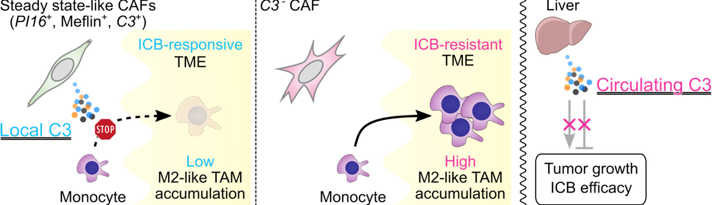
The researchers found that C3 made inside tumor tissue prevents immunosuppressive myeloid cells from entering the tumor microenvironment, the area surrounding and supporting cancer cells. By limiting these cells, local C3 gives the immune system a better opportunity to attack the cancer.
The results identify C3 as a previously unrecognized regulator of cancer immunotherapy — treatment designed to help the immune system detect and destroy cancer cells.
Blood C3 does not drive the response
To determine whether C3 circulating in the blood also affected immunotherapy, the researchers used mice to distinguish between protein produced by the liver and protein made within tumors. Reducing liver-derived C3 by 90% did not weaken the effects of an anti-PD-1 antibody, a drug that helps immune cells attack tumors.
The outcome changed when researchers stopped fibroblasts inside tumors from producing C3. The same immunotherapy became less effective, even though blood levels of C3 fell by only 9%.
“What determined the efficacy of the immunotherapy treatment was not the C3 in the blood, but the local C3 produced at the tumor site. When this C3 breaks down, it forms a fragment called iC3b that stops harmful myeloid cells from entering the tumor. As a result, immunotherapy is more likely to work,” Miyai explained.
Mimicking C3 revives resistant treatment
The researchers next tested whether they could reproduce C3’s protective effect in cancers that resisted immunotherapy. They used a drug that imitates the way C3 blocks myeloid cells from entering tumors.
The combination made previously resistant tumors respond to immunotherapy and significantly prolonged survival in mice. The findings could eventually help identify patients most likely to benefit from immunotherapy and point toward treatment options for tumors that initially resist it.
Patient tumors show the same pattern
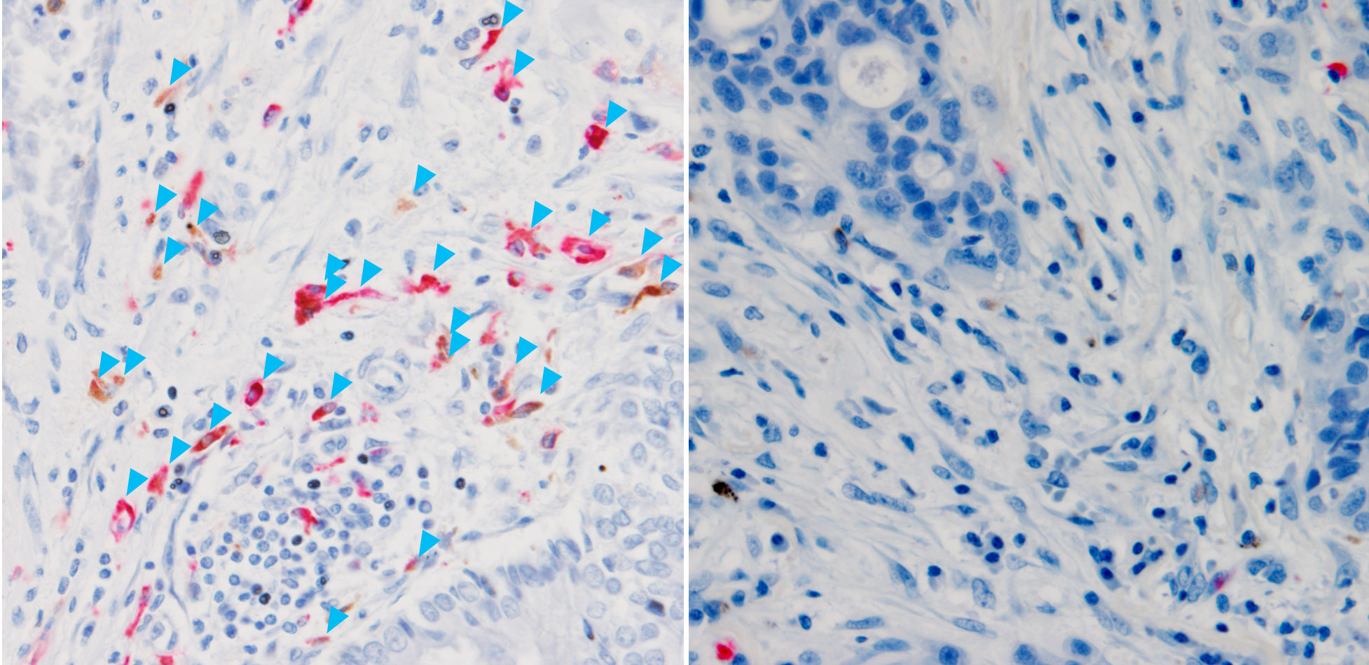
Analysis of lung cancer samples revealed a similar relationship in patients. Those with higher C3 levels in tissue surrounding the cancer had better treatment responses and longer survival. About half of these patients responded to treatment, compared with none of those whose tumors had lower C3 levels. Once again, C3 concentrations in the blood were unrelated to the outcome.
The researchers now plan to test methods for increasing C3 specifically within tumors and determine when such treatment would be most effective. They also believe that studying the protein’s local activity could improve understanding of other processes involving immune regulation, including wound healing and inflammation.
Reference: “Local, but not circulating, complement C3 shapes immune checkpoint blockade efficacy by controlling myeloid cell infiltration” by Yuki Miyai, Yukihiro Shiraki, Ryota Ando, Daisuke Sugiyama, Yoshitaka Sato, Katsuhiro Kato, Naoya Asai, Fuyang Cao, Nobuyoshi Nagao, Kana Tanabe, Masahiro Nakatochi, Tetsunari Hase, Toyofumi Fengshi Chen-Yoshikawa, Tomoko Kobayashi, Shintaro Iwama, Nobutoshi Esaki, Shinji Mii, Hiroshi Arima, Hiroshi Kimura, Masahide Takahashi, Yuichi Ando and Atsushi Enomoto, 15 July 2026, Nature Communications.
DOI: 10.1038/s41467-026-75542-3
This study was supported by funds from the Ministry of Education, Culture, Sports, Science, and Technology of Japan (23K14591, 25K18851, 22H03350, 22H04923, 20H03528, 22H02848, 22K18390), JSMO/KFCR Young Investigator Research Grant, Aichi Cancer Research Foundation, Japan Agency for Medical Research and Development (JP24gm1210009, JP24ama221333), Naito Foundation, Princess Takamatsu Cancer Research Fund, DAIKO Foundation, and Toyoaki Foundation.
Never miss a breakthrough: Join the SciTechDaily newsletter.
Follow us on Google and Google News.