The study findings highlight the crucial impact of sensory experiences on brain development.
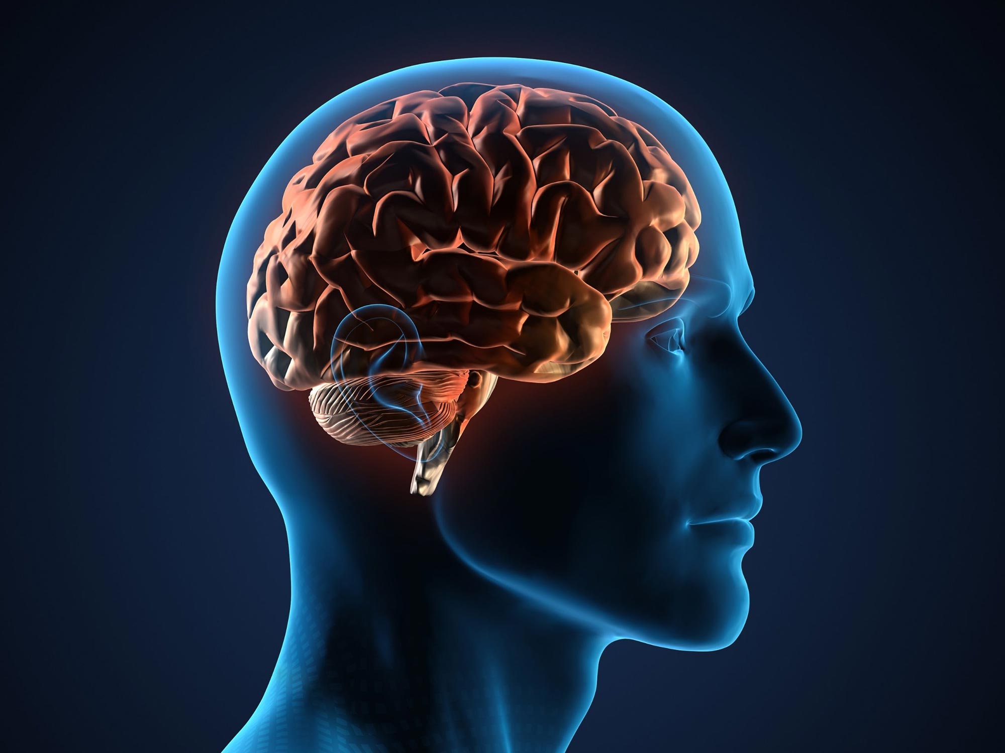
Scientists have long understood that our brains are structured into distinct areas, each dedicated to specific functions. For example, the visual cortex handles what we see, and the motor cortex controls movement. However, the mechanisms of how these regions form—and how their neural building blocks differ—remain a mystery.
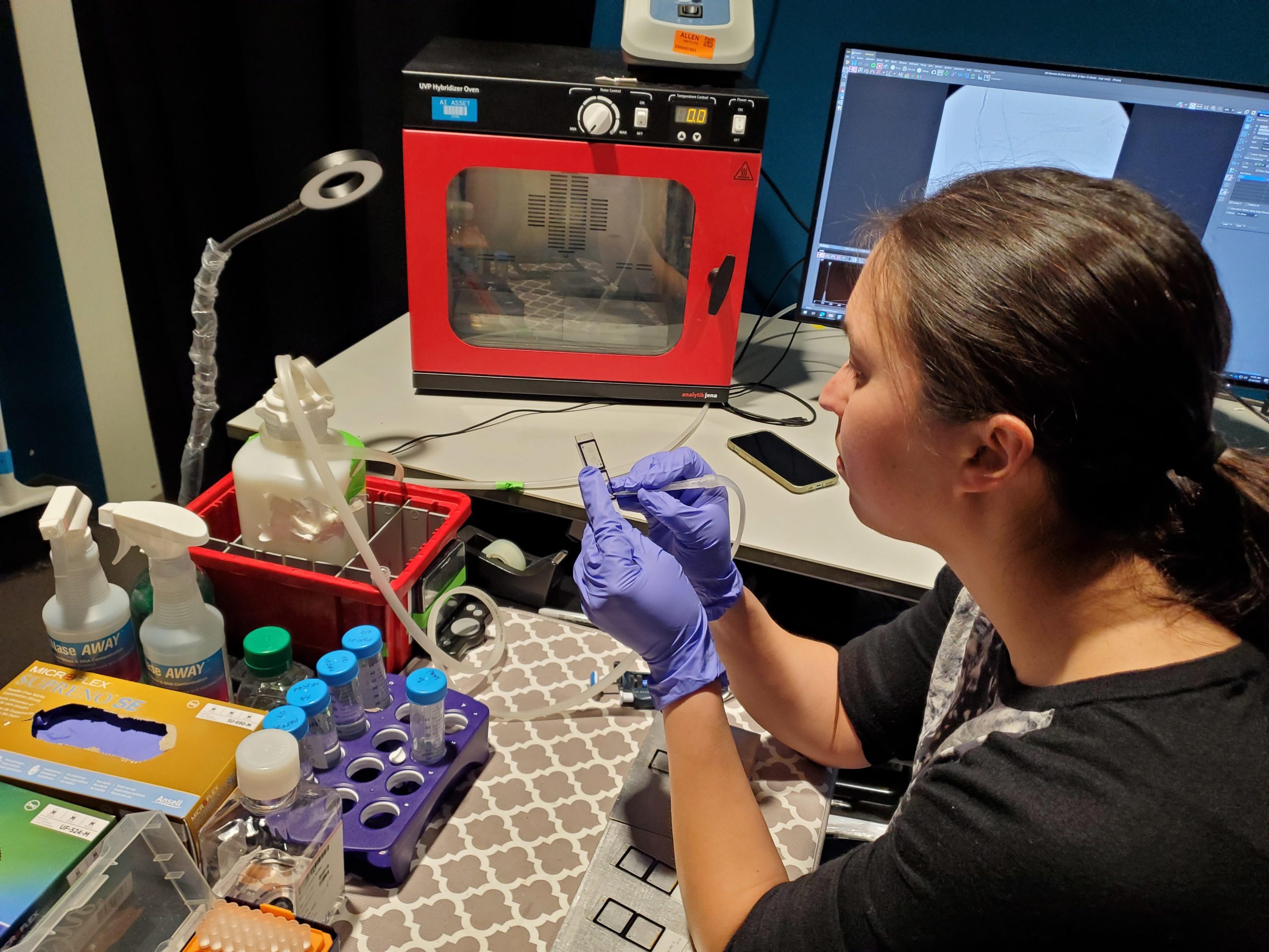
A study published in Nature sheds new light on the brain’s cellular landscape. Researchers at the Allen Institute for Brain Science used an advanced method called BARseq to swiftly classify and map millions of neurons across nine mouse brains. They discovered that while brain regions share the same types of neurons, the specific combination of these cells gives each area a distinct ‘signature,’ akin to a cellular ID card.
The team further explored how sensory inputs influence these cellular signatures. They discovered that mice deprived of sight experienced a major reorganization of cell types within the visual cortex, which blurred the distinctions with neighboring areas. These shifts were not confined to the visual area but occurred across half of cortical regions, though to a lesser extent.
The study underscores the pivotal role of sensory experiences in shaping and maintaining each brain region’s unique cellular identity.
“BARseq lets us see with unprecedented precision how sensory inputs affect brain development,” said Xiaoyin Chen, Ph.D., the study’s co-lead author and an Assistant Investigator at the Allen Institute. “These broad changes illustrate how important vision is in shaping our brains, even at the most basic level.”
A powerful new brain mapping tool
Previously, capturing single-cell data across multiple brains was challenging, said Mara Rue, Ph.D., co-lead author and a Scientist at the Allen Institute. But BARseq is cheaper and less time-consuming than similar mapping technologies, she said, enabling researchers to examine and compare brain-wide molecular architecture across multiple individuals.
BARseq tags individual brain cells with unique RNA ‘barcodes’ to track their connections across the brain. This data, combined with gene expression analysis, allows scientists to pinpoint and identify vast numbers of neurons in tissue slices.
For this study, the research team used BARseq as a standalone method to rapidly analyze gene expression in intact tissue samples. In just three weeks, the researchers mapped more than 9 million cells from eight brains.
The scale and speed of BARseq provides scientists with a powerful new tool to delve deeper into the intricacies of the brain, Chen said.
“BARseq allows us to move beyond mapping what a ‘model’ or ‘standard’ brain looks like and start to use it as a tool to understand how brains change and vary,” Chen said. “With this throughput, we can now ask these questions in a very systematic way, something unthinkable with other techniques.”
Chen and Rue emphasized that the BARseq method is freely available. They hope their study encourages other scientists to use it to investigate the brain’s organizational principles or zoom in on cell types associated with disease.
“This isn’t something that only the big labs can do,” Rue said. “Our study is a proof of principle that BARseq allows a wide range of people in the field to use spatial transcriptomics to answer their own questions.”
Reference: “Whole-cortex in situ sequencing reveals input-dependent area identity” by Xiaoyin Chen, Stephan Fischer, Mara C. P. Rue, Aixin Zhang, Didhiti Mukherjee, Patrick O. Kanold, Jesse Gillis and Anthony M. Zador, 24 April 2024, Nature.
DOI: 10.1038/s41586-024-07221-6
Never miss a breakthrough: Join the SciTechDaily newsletter.
Follow us on Google and Google News.