
EMBL researchers have created a new AI tool that uses a “molecular laser tag” approach to identify cells capable of revealing the earliest origins of cancer.
The human body depends on accurate genetic instructions to keep its cells working properly. Cancer begins to form when these instructions become disrupted. As genetic mistakes build up over time, cells can lose their normal limits on growth and start multiplying in an uncontrolled way. Chromosomal abnormalities – numerical and structural defects in chromosomes – are often one of the earliest changes that push healthy cells toward becoming cancerous.
Researchers in the Korbel Group at EMBL Heidelberg have created a new AI-based tool that gives scientists a way to closely examine how these chromosomal abnormalities develop. The insights gained from this approach may eventually clarify some of the earliest steps that lead to cancer.
“Chromosomal abnormalities are a main driver for particularly aggressive cancers, and they’re highly linked to patient death, metastasis, recurrence, chemotherapy resistance, and fast tumor onset,” said Jan Korbel, senior scientist at EMBL and senior author of the new paper, published in the journal Nature. “We wanted to understand what determines the likelihood that cells undergo such chromosomal alterations, and what’s the rate at which such abnormalities arise when a still normal cell divides.”
The connection between abnormal chromosomes and cancer has been considered for a long time. More than 100 years ago, German scientist Theodor Boveri proposed, based on his microscopy observations, that irregular chromosomal content in cells plays a role in driving cancer formation.
Why Chromosomal Abnormalities Are Hard to Study
However, spotting these abnormalities has long been difficult because only a small number of cells show them at any moment, and many of those cells either die on their own or are removed through natural selection (or are killed off). Researchers traditionally had to look for these cells by hand through a microscope, and they could collect only a few at once for more detailed study.
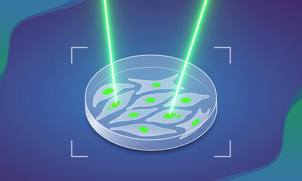
Machine learning-assisted genomics and imaging convergence (MAGIC). Credit: Daniela Velasco/EMBL
Marco Cosenza, Research Scientist in the Korbel Group, hit upon the solution to this problem after working with other teams at EMBL wrestling with similar challenges. He and his collaborators developed a new, autonomous system that combines automated microscopy, single-cell sequencing, and AI, which they named machine learning-assisted genomics and imaging convergence – or MAGIC.
‘Laser tag’ to precisely identify and mark cells
Essentially, MAGIC operates like a fully automated game of laser tag. It spots ‘enemies’, or cells, with a particular kind of visible feature. For this study, the scientists focused on a cellular structure called a ‘micronucleus’. Micronuclei are tiny enclosed compartments inside cells that contain a small portion of the cells’ DNA, broken off from the bulk of the genome. Cells with micronuclei tend to produce new chromosomal abnormalities, which makes them more likely to turn cancerous.
Once cells with micronuclei are detected, the system ‘tags’ them using a laser. For this, the scientists used a photoconvertible dye – a fluorescent molecule that undergoes a chemical transformation if light is shone on it, changing the color of light it emits.
“This project combined a lot of my interests in one,” said Cosenza. “It involves genomics, microscopic imaging, and robotic automation. During the COVID-19-related lockdown in 2020, I could really spend some time on learning and applying AI computer vision technologies to the biological image data we had collected before. Afterwards, we designed experiments to validate it and take it further.”
In practice, MAGIC works like this. First, an automated microscope captures a series of images of a cell sample. Next, a machine learning algorithm, trained on manually annotated datasets of micronuclei-containing cells, scans the images. When the algorithm spots cells with micronuclei, it shares their location with the microscope and instructs it to shine light specifically on those cells, permanently tagging them. The tagged cells can then easily be separated from these still-living cells using methods like flow cytometry, and subsequently be subjected to deeper analysis, e.g. by looking at their cellular genomes.
Scaling Up a Previously Slow Process
By automating the previously labor-intensive, time-consuming, and error-prone process of detecting cells with micronuclei, MAGIC allows scientists to study such cells at a scale and speed previously unheard of. In less than a day, scientists can analyze nearly 100,000 cells using this method.
The team used MAGIC to analyze chromosomal abnormalities in cultured cells originally derived from normal human cells. Their results showed that a little more than 10% of all cell divisions result in spontaneous chromosomal abnormalities of some kind and that this rate nearly doubles when a particular gene – p53, a well-known tumor suppressor – is mutated. The scientists also studied other triggers and contributors to chromosomal abnormality formation, such as the presence and location of double-stranded DNA breaks within a chromosome.
The study involved collaborations across and outside EMBL, with key contributions from the Advanced Light Microscopy Facility (ALMF) and the Pepperkok Team at EMBL Heidelberg, Isidro Cortes-Ciriano’s group at EMBL-EBI, and Andreas Kulozik’s team at the German Cancer Research Centre (DKFZ), which also forms part of the Molecular Medicine Partnership Unit (MMPU) between EMBL and the University of Heidelberg.
MAGIC is a highly versatile and adaptable technique. While the scientists trained it for this study to spot cells that had micronuclei, the algorithm can, in theory, be trained on many different kinds of datasets to detect different cellular features.
“As long as you have a feature that can be discriminated visually from a ‘regular’ cell, you can – thanks to AI – train the system to detect it,” said Korbel, “Our system therefore has potential to advance future discoveries in numerous areas of biology.”
Reference: “Origins of chromosome instability unveiled by coupled imaging and genomics” by Marco Raffaele Cosenza, Alice Gaiatto, Büşra Erarslan Uysal, Álvaro Andrades, Nina Luisa Sautter, Marina Simunovic, Michael Adrian Jendrusch, Sonia Zumalave, Tobias Rausch, Aliaksandr Halavatyi, Eva-Maria Geissen, Joshua Lucas Eigenmann, Thomas Weber, Patrick Hasenfeld, Eva Benito, Catherine Stober, Isidro Cortes-Ciriano, Andreas E. Kulozik, Rainer Pepperkok and Jan O. Korbel, 29 October 2025, Nature.
DOI: 10.1038/s41586-025-09632-5
Never miss a breakthrough: Join the SciTechDaily newsletter.
Follow us on Google and Google News.
5 Comments
Hello sir namaste god bless all family members danke fur god Schone Gruss Rajveer Singh sun of karnail singh batth village kotala bet po chhourian district Ludhiana Punjab India thanks for alls
2 years ago i was told i had
melanoma. several spots that looked suspicious.
5 biopsies with 4 positives.
topical chemo cream for all
but 3 spots.
one was burned off.
the other two spots under-
went an excision.
i did not tell my doctor but
upon getting my positive for
cancer verdict i started to
take 48 mg of ivermectin
each day. i weigh 180 lbs.
after taking 4-12mg tabs
daily for 6 weeks i had my
first excision. it was a funky
mole on my side torso.
a week later, upon starting
the second excision on my
lower back, my doctor said
“IT APPEARS YOUR
BODY IS ATTEMPTING
TO HEAL ITSELF”!
my bloody, raw, seeping
quarter size spot had started
drying out & forming a scab!
from my readings it sounds
like ivermectin stops a cancer cell from being able
to feed itself. it starves it!
i took 4 pills a day for 3 more
weeks after the excisions.
i then went to 1 tab a day &
i continue to take 1 a day.
2 years later no cancer.
no spots. no covid. i have
not had any illness since…
Where did you get ivermectin?
Why did the investigators choose micronuclei, instead of nuclear DNA itself? Moreover did they look at embryonic cells and micronuclei in similar experiments. Though I cannot quote, one school of thought that several Cancers have precursors and number of checks prevent the cells going into mitotic figures?
Do investigators look at epigenetic levels and influence of growth factors on triggering of transcription in similar studies ? p53 has been in genomic science for long time, but since then there so many other tumor suppressor and tumor promoters which have been enumerated!
Astronomers face a similar problem analyzing astronomical data. They might have techniques that could be applied to this problem.