Caltech’s new photoacoustic vector tomography (PAVT) technology enables groundbreaking noninvasive imaging of deep blood vessels and detailed analysis of blood flow dynamics.
A great number of health problems, and consequently the medical treatments for them, involve how blood flows through the body. Heart attacks are caused by restricted blood flow to the heart muscle. Many symptoms of diabetes are the result of damaged blood vessels. Tumors, meanwhile, often promote the growth of new vessels that deliver blood specifically to them. And blood flow is a crucial physiological parameter for measuring brain function.
Because of this, medical professionals want to be able to examine blood vessels and assess their condition, but with many of those vessels buried quite deeply in the body, such an examination can be difficult without exploratory surgery.
New research conducted in the laboratory of Caltech’s Lihong Wang, Bren Professor of Medical Engineering and Electrical Engineering, is now making it possible to image deep blood vessels in humans, and even the blood flowing through them, in a noninvasive way.
Innovative Imaging Technology: PAVT
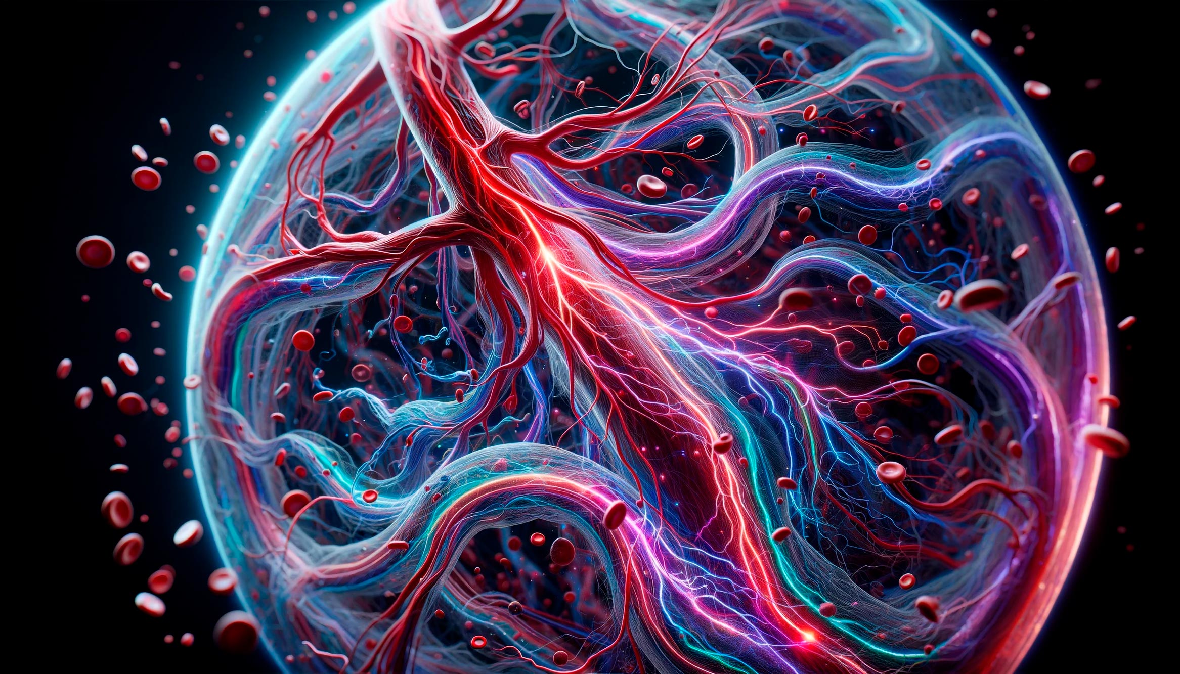
In an article published in the journal Nature Biomedical Engineering, Wang and his colleagues describe this technology, which they call photoacoustic vector tomography, or PAVT. This technology is similar in many ways to Wang’s other photoacoustic imaging technologies, which make use of laser light that is well absorbed by hemoglobin, the oxygen-carrying molecule found in red blood cells.
The energy that the hemoglobin molecules absorb from the laser causes them to vibrate ultrasonically. Those vibrations travel throughout the tissue until they arrive at the skin’s surface, where they are detected by sensors connected to a computer. The computer then creates an image of the features of the tissue, in this case, the blood vessels.
This is not the first time Wang’s lab has shown the ability to image blood vessels using photoacoustic technology, but the new method can image blood flow deeper in the human body than previously possible and shows for the first time not just the presence of blood vessels and their oxygenation status but how the blood is flowing through the vessels.
Breakthrough in Blood Flow Imaging
“Before, we could only show the sizes of blood vessels, concentrations of blood, and oxygen saturations,” says Wang, also the Andrew and Peggy Cherng Medical Engineering Leadership Chair. “Now, we can measure the vector flow, which indicates both flow rate and direction.
Our field has been working on photoacoustic technology for more than 20 years, but nobody predicted anything like this. We surprised ourselves because our field didn’t think this was possible.”
“When I first saw our images of blood flow, I was absolutely amazed,” says Yang Zhang, the lead author and a postdoctoral scholar research associate in medical engineering. “The most exciting part of this work is that we synergized engineering and physiology to overcome a hurdle previously thought to be insurmountable by the field.”
The team is able to see the direction and flow rate because PAVT has such fine resolution that it can make out signals arising from the distribution of red blood cells deep in the body. An algorithm integrated into the system tracks the motion of these distributions and deduces the speed and direction of the flow. It’s kind of like how Google determines how heavy traffic is on a freeway by looking at the speed at which mobile phones are moving in that area.
The researchers hypothesize that their images and videos of human blood flow are facilitated by the heterogenous distribution of red blood cells, which arises, in part, from the way blood vessels are structured throughout the body.

Wang likens the situation in veins to what happens when two rivers with different water qualities, one clear and one muddy, for example, join into one larger stream. At such a confluence, it is not uncommon to see the streams remain unmixed for a long distance even while flowing through the same channel.
A similar phenomenon is seen when two veins carrying blood with differing blood contents (oxygenated and unoxygenated) join together. Even though the blood from those two vessels has joined as a single stream, it will remain unmixed for a while. The PAVT system can distinguish these unmixed patches and track their motion.
And since red blood cells absorb laser light from the PAVT system differently depending on whether they are oxygenated or not, PAVT can also determine how much oxygen the blood in a particular vessel is carrying. “This allows us to quantify oxygen consumption, which is an important measure of metabolism,” Wang adds.
Reference: “Photoacoustic vector tomography for deep haemodynamic imaging” by Yang Zhang, Joshua Olick-Gibson, Anjul Khadria and Lihong V. Wang, 30 November 2023, Nature Biomedical Engineering.
DOI: 10.1038/s41551-023-01148-5
In addition to Wang and Zhang, other co-authors are Joshua Olick-Gibson, medical engineering graduate student, and Anjul Khadria, formerly a Caltech postdoctoral scholar research associate.
Funding for the research was provided by the National Institutes of Health.
Never miss a breakthrough: Join the SciTechDaily newsletter.
Follow us on Google and Google News.