Proteins that encapsulate viruses can be molded into defined shapes using DNA and RNA origami nanostructures.
Bioengineers have discovered a method to customize the size and shape of virus particles. This new approach, which involves merging viral protein building blocks and DNA templates, offers potential applications in the fields of vaccine creation and drug delivery.
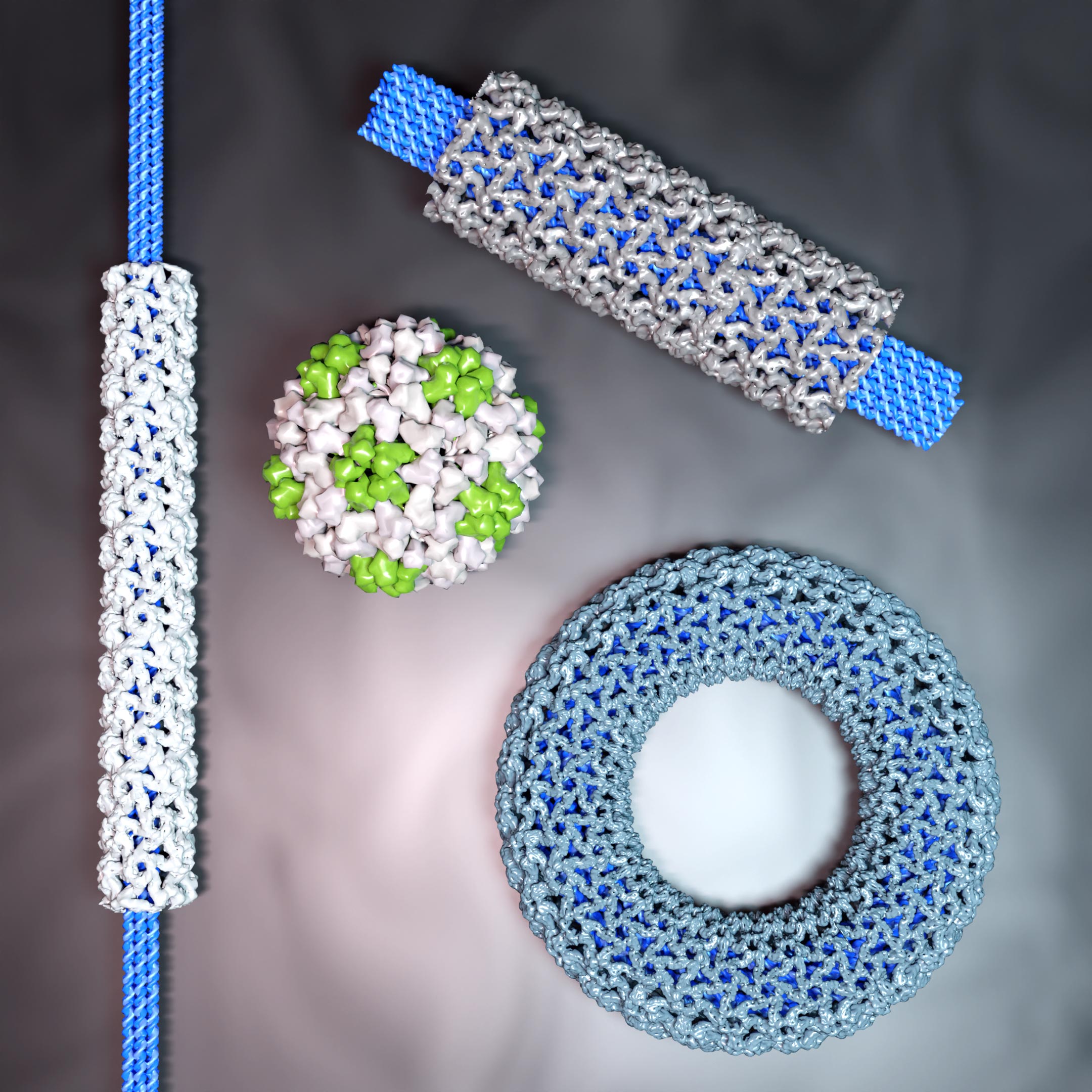
Using Virus Capsid Proteins
Virus capsid proteins, the protective shield for a virus’s genome, can serve as a base for creating meticulously structured protein assemblies. However, their shapes and geometry primarily depend on the virus strain. Reprogramming these assemblies, irrespective of the original viral blueprint, presents a tantalizing possibility in areas such as drug delivery and vaccine development.
The scientific team addressed this challenge by generating a “structured genome” template for the assembly of capsid proteins. They utilized rigid DNA origami structures to prevent deformation of the flexible genome and the formation of unwanted shapes. These structures are tiny in size, ranging from tens to hundreds of nanometers, but entirely made of DNA, which is precisely folded into the desired template shape.
The Role of Electrostatic Interactions
“Our approach is based on electrostatic interactions between the negative charge of the DNA nanostructures and a positively charged domain of the capsid proteins, paired with intrinsic interactions between the single proteins. By altering the amount of protein used, we can fine-tune the number of highly-ordered protein layers, which encapsulate the DNA origami,” says Iris Seitz, lead author and doctoral researcher at Aalto University.
“By using DNA origami as a template, we can direct the capsid proteins into a user-defined size and shape, resulting in assemblies which are well-defined, both in length and diameter. By testing a variety of DNA origami structures, we also learned how the templates’ geometry affected the whole assembly,” Seitz adds.
Cryogenic Electron Microscopy Imaging
“With the help of cryogenic electron microscopy imaging, we were able to visualize the highly ordered proteins upon assembly and, with that, measure even small changes in the geometry of the assembly arising from different templates,” explains professor Juha Huiskonen, a collaborating scientist from the University of Helsinki.
Relevance and Applications
“We have found a simple but effective strategy to (re)direct capsid proteins to a desired shape. Our approach is adaptable and therefore not limited to a single capsid protein type, as we demonstrated with capsid proteins from four different viruses. Additionally, we can tweak our template to be more application-relevant, for instance by integrating RNA into the origami, which could subsequently be translated into useful or site-specific proteins,” explains Aalto professor Mauri Kostiainen, leader of the research project.
Although DNA origami structures are a promising material for interfacing biological systems, they suffer from instability, especially in the presence of DNA-degrading enzymes.
In experiments, however, “we can clearly observe that the protein layer efficiently protects the encapsulated DNA nanostructures from degradation. By combining protection with the functional properties of nucleic acid origami, including the possibility to deliver DNA or messenger RNA together with other cargo molecules, we believe that our approach provides interesting future directions for biomedical engineering,” concludes Kostiainen.
Reference: “DNA-origami-directed virus capsid polymorphism” by Iris Seitz, Sharon Saarinen, Esa-Pekka Kumpula, Donna McNeale, Eduardo Anaya-Plaza, Vili Lampinen, Vesa P. Hytönen, Frank Sainsbury, Jeroen J. L. M. Cornelissen, Veikko Linko, Juha T. Huiskonen and Mauri A. Kostiainen, 17 July 2023, Nature Nanotechnology.
DOI: 10.1038/s41565-023-01443-x
This work was conducted jointly at Aalto University (Finland) with researchers from the University of Helsinki (Finland), Griffith University (Australia), Tampere University (Finland) and University of Twente (The Netherlands).
Never miss a breakthrough: Join the SciTechDaily newsletter.
Follow us on Google and Google News.