A Hereditary Condition That Affects the Brain’s Neurons
Huntington’s disease is a genetic neurodegenerative condition that results in motor, cognitive, and psychiatric impairments in those afflicted. Grasping the changes in the brain’s neural pathways in this disorder is crucial for developing therapeutic strategies. The disease has been linked to the malfunctioning of certain neuronal pathways, particularly the corticostriatal circuitry, in patients.
Now, a study published in the Journal of Neuroscience has discovered further alterations in other neural circuits using mouse models to study this pathology, which profoundly impacts the patients’ lives.
The study was led by Mercè Masana, lecturer at the Faculty of Medicine and Health Sciences of the University of Barcelona and member of the UB Institute of Neurosciences (UBneuro), the August Pi i Sunyer Biomedical Research Institute (IDIBAPS), and the Biomedical Research Networking Center on Neurodegenerative Diseases (CIBERNED). The study, whose first author is the researcher Sara Conde Berriozabal, includes the participation of the experts Jordi Alberch, Manuel José Rodríguez, and Guadalupe Soria (UB, UBneuro, IDIBAPS), among others. The study has been carried out with the support from the UB Scientific and Technological Centers (CCiTUB) and the IDIBAPS Magnetic Resonance Imaging Unit.
An Inherited Disorder That Affects Neurons in the Brain
Huntington’s disease is a rare, inherited disease that usually manifests in adults aged between 35 and 50, although there are also some juvenile forms of the disease. It is caused by a mutation in the gene called IT15 or HTT, which codes for huntingtin protein (HTT). Historically, the motor disorder that was most commonly associated with the disorder was chorea —which causes abnormal, involuntary movements— but there are also other non-motor disorders that often appear earlier.
This disorder is associated with dysfunction of corticobasal circuits in the brain. In a previous study, published in the journal eLife (2020), the team characterized one of the neural circuits involved in the development of the disease in animal models: the connection from the secondary motor cortex (M2) to the dorsolateral striatum nucleus (DSL).
In patients, the most affected brain area from the beginning of the disease is the premotor cortex —the M2 cortex in mice— which is involved in cognitive functions and perceptual processes. In the case of animal models, the M2 is associated with motor learning deficits. Moreover, this cortical area is known to be able to project neuronal axons to various brain regions beyond the striatum nucleus.

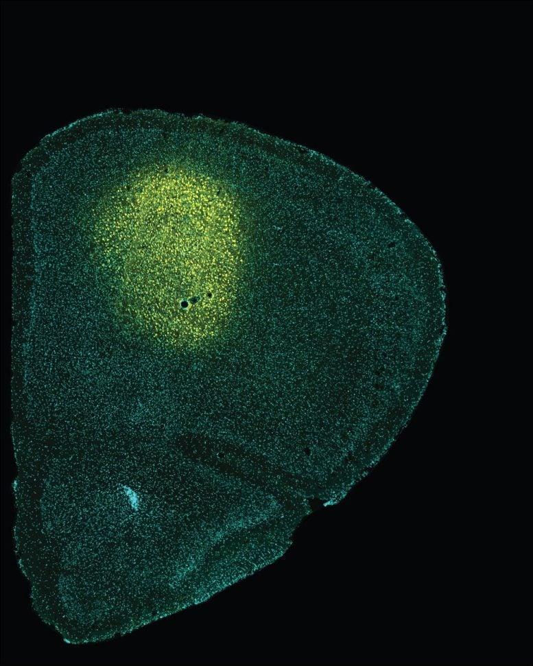
Now, this study has identified for the first time that the M2 cortex sends different axonal projections to another anatomical structure in the brain —the superior colliculus (SC). These projections are deeply impaired and could be linked to the disease symptomatology.
As part of the study, the functional magnetic resonance imaging revealed the reduced functional connectivity between the left M2 cortex and all the brain regions analyzed in mice models of the disease. By applying other innovative methodologies to monitor and modulate neural activity —ontogeny, electrophysiology, photometry, and chemogenetics— the team discovered that the lack of M2 cortex activity could be responsible for the altered responses in Huntington’s disease.
Understanding the Alterations in Brain Circuitry
Identifying the different alterations and functions of the M2 cortex circuitry —beyond the cortico-striatal pathway— provides data that are crucial to further analyze the symptoms of Huntington’s disease and other neurodegenerative pathologies (Parkinson’s disease, etc.). Also, a deeper understanding of the role of the superior colliculus and its neural circuits —involved in many neurological disorders such as Huntington’s— may provide new insights into delaying the onset and severity of the symptoms in motor disorders.
Reference: “M2 Cortex Circuitry and Sensory-Induced Behavioral Alterations in Huntington’s Disease: Role of Superior Colliculus” by Sara Conde-Berriozabal, Lia García-Gilabert, Esther García-García, Laia Sitjà-Roqueta, Xavier López-Gil, Emma Muñoz-Moreno, Mehdi Boutagouga Boudjadja, Guadalupe Soria, Manuel J Rodríguez, Jordi Alberch and Mercè Masana, 3 May 2023, Journal of Neuroscience.
DOI: 10.1523/JNEUROSCI.1172-22.2023
Never miss a breakthrough: Join the SciTechDaily newsletter.
Follow us on Google and Google News.