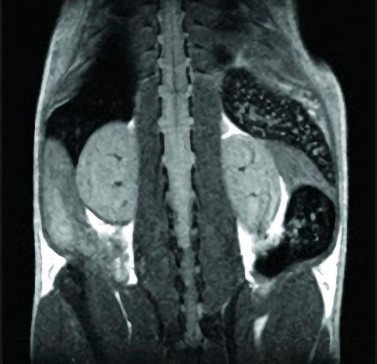
Non-invasive visualization of a mouse by Magnetic Resonance Imaging with a 4.7-Tesla microimaging system. Credit: Wenxian Fu
While working with mice, researchers were able to use a new magnetic nanoparticle-based MRI technique to predict Type-1 diabetes and were able to leverage this diagnostic information to prevent diabetes in mice predisposed to it. They believe this technique has the potential to be a useful tool for performing long-term, longitudinal studies of the progression of diabetes in humans.
Researchers from Harvard Medical School and Massachusetts General Hospital have developed a magnetic nanoparticle-based MRI technique for predicting whether—and when—subjects with a genetic predisposition for diabetes will develop the disease. While done initially in mice, preliminary data show that the platform can be used in people as well, so far to distinguish patients that do or do not have pancreas inflammation.
“This research is about predicting Type-1 diabetes, and using that predictive power to figure out what is different between those who get it and those who don’t get it,” said Diane Mathis, Morton Grove-Rasmussen Professor of Immunohematology in the Department of Microbiology and Immunobiology and, along with Christophe Benoist, Morton Grove-Rasmussen Professor of Immunohematology, co-senior author of the paper. The results were published online in Nature Immunology on February 26, 2012.
According to first author Wenxian Fu, a research fellow in the Mathis-Benoist lab, the group was surprised that the diagnostic window—from six to 10 weeks of age—was so early, and so brief. This shows that the progression of the disease, at least in this animal model, is determined very early in life, and that diabetes does not require an additional trigger such as a secondary infection or environmental stress, as some theoretical conceptions of diabetes have proposed.
What’s more, the researchers were able to leverage this diagnostic information to prevent diabetes in mice predisposed to it.
Invisible differences
Even within a population of genetically identical mice, all of which are predisposed to develop Type 1 diabetes, there is great variability in the course of the disease. Some 30 percent never develop diabetes; among those that do, it might develop in the first months of life, or not until six months.
Until now, when researchers wanted to study the fundamental biochemical and biophysical development of diabetes, they had to assume that all of the subjects in their study were at similar stages in the progression of the disease, even though, in reality, nearly a third of the animals would never even develop the disease—and there was no way to know which animals were part of that group. This uncertainty adds significant noise to the data.
Using the techniques developed by Mathis and Benoist’s group in a ten-year collaboration with the laboratory of Ralph Weissleder, HMS professor of systems biology and radiology at MGH, Fu discovered that he could not only predict which mice would develop diabetes, but how soon.
“There’s a nice correlation between the intensity of the signal and how quickly they will develop diabetes,” Fu said.
The researchers are hopeful that the scanning method will allow scientists to study with greater accuracy and uniformity the developmental stages of this disease.
Setting their sights on inflammation
The mice in the study were injected with magnetic nanoparticles designed to accumulate only in inflamed tissues, where they are then visible by MRI. The team performed full-body scans of the subjects, but focused their attention on inflammation in the pancreas.
Fu and collaborators performed periodic scans over the course of 18 weeks, then waited until the mice were 40 weeks old to monitor for signs of diabetes. Looking at the data retrospectively, they saw a clear pattern: In mice that developed diabetes, there was increased inflammation in the pancreas between the ages of six and 10 weeks. Outside of this window, there was no difference in MRI signal between the two groups.
“It’s important to note that we could not have made this discovery without a non-invasive scanning technique, because otherwise the mice would need to be sacrificed before knowing whether they go on to develop the disease,” said, Benoist.
Once they could predict which mice would and would not become diabetic, they performed biochemical and gene expression analyses of the mice, separating cohorts of diabetic and non-diabetic mice. They identified a number of markers that correlate with diabetes development.
The researchers speculate that [in many of these cases,] increased expression of a particular gene in non-diabetic subjects conveys protection from diabetes. One of the pathways they examined was a receptor called CRIg, a molecule involved in various innate immune system activities. Injecting engineered CRIg-related molecules into mice predisposed to diabetes inhibited disease development, demonstrating the important role this pathway plays in the disease.
In an earlier study, the collaborative team confirmed that the same scanning techniques could be used to measure pancreatic inflammation in human subjects, and were able to separate non-diabetics from diabetics. The great value of this new ability is that it will permit a rapid assessment of the influence of drugs designed to clear the pancreatic inflammation. “We could use our MRI technique to get real-time data on the effectiveness of new drug therapies,” Mathis said. “The way we work now, we have to wait and wait to see if therapies are having any benefit to the patients.”
Mathis said that their technique also has the potential to be a useful tool for performing long term, longitudinal studies of the progression of diabetes in humans. Current methods of imaging the pancreas, such as high-radiation PET scans, are too dangerous to use for repeated scans.
Reference: “Early window of diabetes determinism in NOD mice, dependent on the complement receptor CRIg, identified by noninvasive imaging” by Wenxian Fu, Gregory Wojtkiewicz, Ralph Weissleder, Christophe Benoist and Diane Mathis, 26 February 2012, Nature Immunology.
DOI: 10.1038/ni.2233
This research was supported by the National Institutes of Health, the Juvenile Diabetes Research Foundation, and the American Diabetes Association.


“The researchers speculate” so this is still in infant stage research and we don’t have anything concrete so far…